- Home
- About
- Promotions
-
Products
-
Elisa Kits
- |
-
Primary antibodies
- |
-
Secondary antibodies
- |
-
Proteins
- |
-
IHC reagents
- |
-
WB reagents
- PonceauS Staining Solution
- PBST Washing Buffer, 10X
- 1.5M Tris-HCl Buffer, pH8.8
- 1M Tris-HCl Buffer, pH6.8
- 10% SDS Solution
- Prestained Protein Marker
- TBST Washing Buffer, 10X
- SDS PAGE Loading Buffer, 5X
- Stripping Buffered Solution
- Tris Buffer, pH7.4, 10X
- Total Protein Extraction Kit
- Running Buffer, 10X
- Transfer Buffer, 10X
- 30% Acr-Bis(29:1) Solution
- Tris电泳液速溶颗粒
- PBS(1X, premixed powder)
- TBS(1X, premixed powder)
- 快速封闭液
- 转膜液速溶颗粒
- Chemical reagents
- News
- Distributor
- Resources
- Contact
- Home
- >
- Info
- >
- EMAL4 rabbit pAb
- >
- Go Back
EMAL4 rabbit pAb
- Catalog No.:YT7154
- Applications:WB
- Reactivity:Human;Mouse
- Fields:
- >>Pathways in cancer;>>Non-small cell lung cancer;>>PD-L1 expression and PD-1 checkpoint pathway in cancer
- Gene Name:
- EML4 C2orf2 EMAPL4
- Immunogen:
- Synthesized peptide derived from human EMAL4 AA range: 398-448
- Specificity:
- This antibody detects endogenous levels of EMAL4 at Human/Mouse
- Formulation:
- Liquid in PBS containing 50% glycerol, 0.5% BSA and 0.02% sodium azide.
- Source:
- Polyclonal, Rabbit,IgG
- Purification:
- The antibody was affinity-purified from rabbit antiserum by affinity-chromatography using epitope-specific immunogen.
- Storage Stability:
- -15°C to -25°C/1 year(Do not lower than -25°C)
- Molecular Weight(Da):
- 108kD
- Background:
- This gene is a member of the echinoderm microtubule associated protein-like family. The encoded WD-repeat protein may be involved in microtubule formation. Abnormal fusion of parts of this gene with portions of the anaplastic lymphoma receptor tyrosine kinase gene, which generates EML4-ALK fusion transcripts, is one of the primary mutations associated with non-small cell lung cancer. Alternative splicing of this gene results in two transcript variants. [provided by RefSeq, Jan 2015],
- Function:
- developmental stage:Strongly overexpressed during mitosis.,function:May modify the assembly dynamics of microtubules, such that microtubules are slightly longer, but more dynamic.,similarity:Belongs to the WD repeat EMAP family.,similarity:Contains 9 WD repeats.,
- Subcellular Location:
- Cytoplasm, cytoskeleton . Cytoplasm . Cytoplasm, cytoskeleton, spindle . Cytoplasm, cytoskeleton, microtubule organizing center . Midbody . Localizes to microtubules (MTs) during interphase with a significantly reduced affinity for MTs during mitosis. .
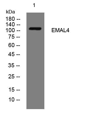
- Western blot analysis of lysates from Jurkat cells, primary antibody was diluted at 1:1000, 4°over night


