ASCC3 rabbit pAb
- Catalog No.:YT6897
- Applications:WB
- Reactivity:Human;Mouse;Rat
- Target:
- ASCC3
- Gene Name:
- ASCC3 HELIC1
- Protein Name:
- ASCC3
- Human Gene Id:
- 10973
- Human Swiss Prot No:
- Q8N3C0
- Mouse Gene Id:
- 77987
- Mouse Swiss Prot No:
- E9PZJ8
- Rat Swiss Prot No:
- F1LPQ2
- Immunogen:
- Synthesized peptide derived from human ASCC3 AA range: 2053-2103
- Specificity:
- This antibody detects endogenous levels of ASCC3 at Human/Mouse/Rat
- Formulation:
- Liquid in PBS containing 50% glycerol, 0.5% BSA and 0.02% sodium azide.
- Source:
- Polyclonal, Rabbit,IgG
- Dilution:
- WB 1:500-2000
- Purification:
- The antibody was affinity-purified from rabbit antiserum by affinity-chromatography using epitope-specific immunogen.
- Concentration:
- 1 mg/ml
- Storage Stability:
- -15°C to -25°C/1 year(Do not lower than -25°C)
- Molecular Weight(Da):
- 242kD
- Background:
- This gene encodes a protein that belongs to a family of helicases that are involved in the ATP-dependent unwinding of nucleic acid duplexes. The encoded protein is the largest subunit of the activating signal cointegrator 1 complex that is involved in DNA repair and resistance to alkylation damage. Alternate splicing results in multiple transcript variants. [provided by RefSeq, Sep 2013],
- Function:
- function:Enhances NF-kappa-B, SRF and AP1 transactivation.,similarity:Belongs to the helicase family.,similarity:Contains 2 helicase ATP-binding domains.,similarity:Contains 2 helicase C-terminal domains.,similarity:Contains 3 SEC63 domains.,subunit:Part of TRIP4 complex, that contains ASCC1, ASCC2 and ASCC3.,tissue specificity:Ubiquitous.,
- Subcellular Location:
- Nucleus . Nucleus speckle . Cytoplasm, cytosol . Colocalizes with ALKBH3 and ASCC2 in nuclear foci when cells have been exposed to alkylating agents that cause DNA damage. .
- Expression:
- Ubiquitous.
- June 19-2018
- WESTERN IMMUNOBLOTTING PROTOCOL
- June 19-2018
- IMMUNOHISTOCHEMISTRY-PARAFFIN PROTOCOL
- June 19-2018
- IMMUNOFLUORESCENCE PROTOCOL
- September 08-2020
- FLOW-CYTOMEYRT-PROTOCOL
- May 20-2022
- Cell-Based ELISA│解您多样本WB检测之困扰
- July 13-2018
- CELL-BASED-ELISA-PROTOCOL-FOR-ACETYL-PROTEIN
- July 13-2018
- CELL-BASED-ELISA-PROTOCOL-FOR-PHOSPHO-PROTEIN
- July 13-2018
- Antibody-FAQs
- Products Images
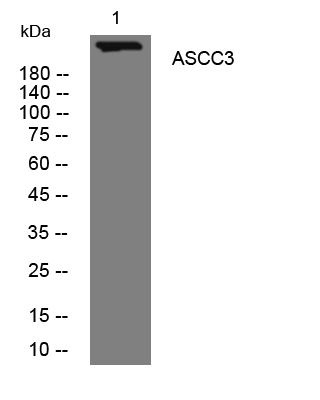
- Western blot analysis of lysates from K562 cells, primary antibody was diluted at 1:1000, 4°over night


