- Home
- About
- Promotions
-
Products
-
Elisa Kits
- |
-
Primary antibodies
- |
-
Secondary antibodies
- |
-
Proteins
- |
-
IHC reagents
- |
-
WB reagents
- PonceauS Staining Solution
- PBST Washing Buffer, 10X
- 1.5M Tris-HCl Buffer, pH8.8
- 1M Tris-HCl Buffer, pH6.8
- 10% SDS Solution
- Prestained Protein Marker
- TBST Washing Buffer, 10X
- SDS PAGE Loading Buffer, 5X
- Stripping Buffered Solution
- Tris Buffer, pH7.4, 10X
- Total Protein Extraction Kit
- Running Buffer, 10X
- Transfer Buffer, 10X
- 30% Acr-Bis(29:1) Solution
- Tris电泳液速溶颗粒
- PBS(1X, premixed powder)
- TBS(1X, premixed powder)
- 快速封闭液
- 转膜液速溶颗粒
- Chemical reagents
- News
- Distributor
- Resources
- Contact
- Home
- >
- Info
- >
- F111A rabbit pAb
- >
- Go Back
F111A rabbit pAb
- Catalog No.:YT6701
- Applications:WB
- Reactivity:Human;Mouse
- Gene Name:
- FAM111A KIAA1895
- Immunogen:
- Synthesized peptide derived from human F111A AA range: 264-314
- Specificity:
- This antibody detects endogenous levels of F111A at Human/Mouse
- Formulation:
- Liquid in PBS containing 50% glycerol, 0.5% BSA and 0.02% sodium azide.
- Source:
- Polyclonal, Rabbit,IgG
- Purification:
- The antibody was affinity-purified from rabbit antiserum by affinity-chromatography using epitope-specific immunogen.
- Storage Stability:
- -15°C to -25°C/1 year(Do not lower than -25°C)
- Molecular Weight(Da):
- 67kD
- Background:
- The protein encoded by this gene is cell-cycle regulated, and has nuclear localization. The C-terminal half of the protein shares homology with trypsin-like peptidases and it contains a PCNA-interacting peptide (PIP) box, that is necessary for its co-localization with proliferating cell nuclear antigen (PCNA). Reduced expression of this gene resulted in DNA replication defects, consistent with the demonstrated role for this gene in Simian Virus 40 (SV40) viral replication. Mutations in this gene have been associated with Kenny-Caffey syndrome (KCS) type 2 and the more severe osteocraniostenosis (OCS, also known as Gracile Bone Dysplasia), both characterized by short stature, hypoparathyroidism, bone development abnormalities, and hypocalcemia. Alternative splicing results in multiple transcript variants. [provided by RefSeq, Aug 2015],
- Function:
- similarity:Belongs to the FAM111 family.,
- Subcellular Location:
- Nucleus . Chromosome . Cytoplasm . Mainly localizes to nucleus: colocalizes with PCNA on replication sites. .
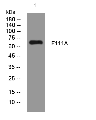
- Western blot analysis of lysates from THP-1 cells, primary antibody was diluted at 1:1000, 4°over night


