- Target:
- RIPK4
- Gene Name:
- RIPK4 ANKRD3 DIK
- Protein Name:
- RIPK4
- Human Swiss Prot No:
- P57078
- Mouse Gene Id:
- 72388
- Mouse Swiss Prot No:
- Q9ERK0
- Immunogen:
- Synthesized peptide derived from human RIPK4 AA range: 280-330
- Specificity:
- This antibody detects endogenous levels of RIPK4 at Human/Mouse
- Formulation:
- Liquid in PBS containing 50% glycerol, 0.5% BSA and 0.02% sodium azide.
- Source:
- Polyclonal, Rabbit,IgG
- Dilution:
- WB 1:500-2000
- Purification:
- The antibody was affinity-purified from rabbit antiserum by affinity-chromatography using epitope-specific immunogen.
- Concentration:
- 1 mg/ml
- Storage Stability:
- -15°C to -25°C/1 year(Do not lower than -25°C)
- Molecular Weight(Da):
- 92kD
- Background:
- The protein encoded by this gene is a serine/threonine protein kinase that interacts with protein kinase C-delta. The encoded protein can also activate NFkappaB and is required for keratinocyte differentiation. This kinase undergoes autophosphorylation. [provided by RefSeq, Jul 2008],
- Function:
- catalytic activity:ATP + a protein = ADP + a phosphoprotein.,function:Plays a role in NF-kappa B activation.,PTM:May be phosphorylated by MAP3K2 and MAP3K3.,PTM:Proteolytically cleaved by during Fas-induced apoptosis. Cleavage at Asp-388 and Asp-426.,similarity:Belongs to the protein kinase superfamily.,similarity:Belongs to the protein kinase superfamily. TKL Ser/Thr protein kinase family.,similarity:Contains 1 protein kinase domain.,similarity:Contains 10 ANK repeats.,subunit:Interacts with PRKCB (By similarity). Interacts with TRAF1, TRAF2, TRAF3 and TRAF5.,
- Subcellular Location:
- Cytoplasm. Membrane ; Peripheral membrane protein .
- Expression:
- Expressed in hair follicles and skin.
Downregulation of RIPK4 Expression Inhibits Epithelial-Mesenchymal Transition in Ovarian Cancer through IL-6. Journal of Immunology Research J Immunol Res. 2021;2021:8875450 WB Human 1 : 1000 SKOV3 cell
- June 19-2018
- WESTERN IMMUNOBLOTTING PROTOCOL
- June 19-2018
- IMMUNOHISTOCHEMISTRY-PARAFFIN PROTOCOL
- June 19-2018
- IMMUNOFLUORESCENCE PROTOCOL
- September 08-2020
- FLOW-CYTOMEYRT-PROTOCOL
- May 20-2022
- Cell-Based ELISA│解您多样本WB检测之困扰
- July 13-2018
- CELL-BASED-ELISA-PROTOCOL-FOR-ACETYL-PROTEIN
- July 13-2018
- CELL-BASED-ELISA-PROTOCOL-FOR-PHOSPHO-PROTEIN
- July 13-2018
- Antibody-FAQs
- Products Images
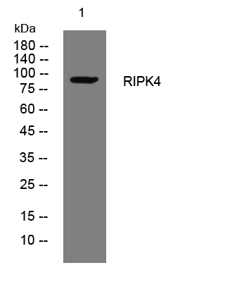
- Western blot analysis of lysates from HuvEc cells, primary antibody was diluted at 1:1000, 4°over night


