PIGL rabbit pAb
- Catalog No.:YT6358
- Applications:WB;IF;ELISA;IHC
- Reactivity:Human;Mouse;Rat
- Target:
- PIGL
- Fields:
- >>Glycosylphosphatidylinositol (GPI)-anchor biosynthesis;>>Metabolic pathways
- Gene Name:
- PIGL
- Protein Name:
- PIGL
- Human Gene Id:
- 9487
- Human Swiss Prot No:
- Q9Y2B2
- Mouse Gene Id:
- 327942
- Mouse Swiss Prot No:
- Q5SX19
- Rat Gene Id:
- 192263
- Rat Swiss Prot No:
- O35790
- Immunogen:
- Synthesized peptide derived from human PIGL AA range: 77-127
- Specificity:
- This antibody detects endogenous levels of PIGL at Human/Mouse/Rat
- Formulation:
- Liquid in PBS containing 50% glycerol, 0.5% BSA and 0.02% sodium azide.
- Source:
- Polyclonal, Rabbit,IgG
- Dilution:
- WB 1:500-2000; IF ICC 1:50-200;ELISA 1:2000-20000;IHC 1:50-200
- Purification:
- The antibody was affinity-purified from rabbit antiserum by affinity-chromatography using epitope-specific immunogen.
- Concentration:
- 1 mg/ml
- Storage Stability:
- -15°C to -25°C/1 year(Do not lower than -25°C)
- Molecular Weight(Da):
- 28kD
- Background:
- This gene encodes an enzyme that catalyzes the second step of glycosylphosphatidylinositol (GPI) biosynthesis, which is the de-N-acetylation of N-acetylglucosaminylphosphatidylinositol (GlcNAc-PI). Study of a similar rat enzyme suggests that this protein localizes to the endoplasmic reticulum. [provided by RefSeq, Jul 2008],
- Function:
- catalytic activity:6-(N-acetyl-D-glucosaminyl)-1-phosphatidyl-1D-myo-inositol + H(2)O = 6-(alpha-D-glucosaminyl)-1-phosphatidyl-1D-myo-inositol + acetate.,function:Involved in the second step of GPI biosynthesis. De-N-acetylation of N-acetylglucosaminyl-phosphatidylinositol.,pathway:Glycolipid biosynthesis; glycosylphosphatidylinositol-anchor biosynthesis.,similarity:Belongs to the PIGL family.,
- Subcellular Location:
- Endoplasmic reticulum membrane ; Single-pass membrane protein .
- June 19-2018
- WESTERN IMMUNOBLOTTING PROTOCOL
- June 19-2018
- IMMUNOHISTOCHEMISTRY-PARAFFIN PROTOCOL
- June 19-2018
- IMMUNOFLUORESCENCE PROTOCOL
- September 08-2020
- FLOW-CYTOMEYRT-PROTOCOL
- May 20-2022
- Cell-Based ELISA│解您多样本WB检测之困扰
- July 13-2018
- CELL-BASED-ELISA-PROTOCOL-FOR-ACETYL-PROTEIN
- July 13-2018
- CELL-BASED-ELISA-PROTOCOL-FOR-PHOSPHO-PROTEIN
- July 13-2018
- Antibody-FAQs
- Products Images
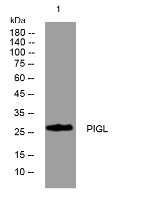
- Western blot analysis of lysates from HpeG2 cells, primary antibody was diluted at 1:1000, 4°over night
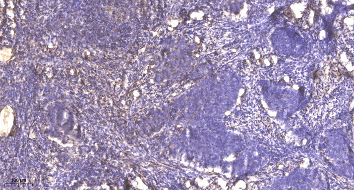
- Immunohistochemical analysis of paraffin-embedded human cervical carcinoma. 1, Antibody was diluted at 1:200(4° overnight). 2, Tris-EDTA,pH9.0 was used for antigen retrieval. 3,Secondary antibody was diluted at 1:200(room temperature, 45min).


