Coronin 1A Polyclonal Antibody
- Catalog No.:YT1061
- Applications:WB;IHC;IF;ELISA
- Reactivity:Human;Mouse;Rat
- Target:
- Coronin 1A
- Fields:
- >>Phagosome;>>Tuberculosis
- Gene Name:
- CORO1A
- Protein Name:
- Coronin-1A
- Human Gene Id:
- 11151
- Human Swiss Prot No:
- P31146
- Mouse Gene Id:
- 12721
- Mouse Swiss Prot No:
- O89053
- Rat Gene Id:
- 155151
- Rat Swiss Prot No:
- Q91ZN1
- Immunogen:
- Synthesized peptide derived from Coronin 1A . at AA range: 150-230
- Specificity:
- Coronin 1A Polyclonal Antibody detects endogenous levels of Coronin 1A protein.
- Formulation:
- Liquid in PBS containing 50% glycerol, 0.5% BSA and 0.02% sodium azide.
- Source:
- Polyclonal, Rabbit,IgG
- Dilution:
- WB 1:500 - 1:2000. IHC 1:100 - 1:300. ELISA: 1:40000.. IF 1:50-200
- Purification:
- The antibody was affinity-purified from rabbit antiserum by affinity-chromatography using epitope-specific immunogen.
- Concentration:
- 1 mg/ml
- Storage Stability:
- -15°C to -25°C/1 year(Do not lower than -25°C)
- Other Name:
- CORO1A;CORO1;Coronin-1A;Coronin-like protein A;Clipin-A;Coronin-like protein p57;Tryptophan aspartate-containing coat protein;TACO
- Observed Band(KD):
- 51kD
- Background:
- This gene encodes a member of the WD repeat protein family. WD repeats are minimally conserved regions of approximately 40 amino acids typically bracketed by gly-his and trp-asp (GH-WD), which may facilitate formation of heterotrimeric or multiprotein complexes. Members of this family are involved in a variety of cellular processes, including cell cycle progression, signal transduction, apoptosis, and gene regulation. Alternative splicing results in multiple transcript variants. A related pseudogene has been defined on chromosome 16. [provided by RefSeq, Sep 2010],
- Function:
- function:May be a crucial component of the cytoskeleton of highly motile cells, functioning both in the invagination of large pieces of plasma membrane, as well as in forming protrusions of the plasma membrane involved in cell locomotion. In mycobacteria-infected cells, its retention on the phagosomal membrane prevents fusion between phagosomes and lysosomes.,similarity:Belongs to the WD repeat coronin family.,similarity:Contains 5 WD repeats.,subcellular location:In non-infected macrophages, associated with the cortical microtubule network. In mycobacteria-infected macrophages, becomes progressively relocalized and retained around the mycobacterial phagosomes. Retention on the phagosomal membrane is strictly dependent on mycobacterial viability and not due to impaired acidification.,subunit:Binds actin.,tissue specificity:Expressed in brain, thymus, spleen, bone marrow and lymph node. L
- Subcellular Location:
- Cytoplasm, cytoskeleton . Cytoplasm, cell cortex . Cytoplasmic vesicle, phagosome membrane . In non-infected macrophages, associated with the cortical microtubule network. In mycobacteria-infected macrophages, becomes progressively relocalized and retained around the mycobacterial phagosomes. Retention on the phagosomal membrane is strictly dependent on mycobacterial viability and not due to impaired acidification (By similarity). .
- Expression:
- Expressed in brain, thymus, spleen, bone marrow and lymph node. Low in lung and gut.
- June 19-2018
- WESTERN IMMUNOBLOTTING PROTOCOL
- June 19-2018
- IMMUNOHISTOCHEMISTRY-PARAFFIN PROTOCOL
- June 19-2018
- IMMUNOFLUORESCENCE PROTOCOL
- September 08-2020
- FLOW-CYTOMEYRT-PROTOCOL
- May 20-2022
- Cell-Based ELISA│解您多样本WB检测之困扰
- July 13-2018
- CELL-BASED-ELISA-PROTOCOL-FOR-ACETYL-PROTEIN
- July 13-2018
- CELL-BASED-ELISA-PROTOCOL-FOR-PHOSPHO-PROTEIN
- July 13-2018
- Antibody-FAQs
- Products Images
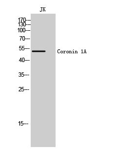
- Western Blot analysis of JK cells using Coronin 1A Polyclonal Antibody
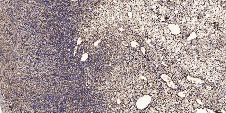
- Immunohistochemical analysis of paraffin-embedded human oophoroma. 1, Antibody was diluted at 1:200(4° overnight). 2, Tris-EDTA,pH9.0 was used for antigen retrieval. 3,Secondary antibody was diluted at 1:200(room temperature, 45min).


