c-Fos Polyclonal Antibody
- Catalog No.:YT0887
- Applications:IF;WB;IHC;ELISA
- Reactivity:Human;Mouse;Rat
- Target:
- c-Fos
- Fields:
- >>Endocrine resistance;>>MAPK signaling pathway;>>cAMP signaling pathway;>>Apoptosis;>>Osteoclast differentiation;>>Toll-like receptor signaling pathway;>>IL-17 signaling pathway;>>Th1 and Th2 cell differentiation;>>Th17 cell differentiation;>>T cell receptor signaling pathway;>>B cell receptor signaling pathway;>>TNF signaling pathway;>>Circadian entrainment;>>Cholinergic synapse;>>Dopaminergic synapse;>>Estrogen signaling pathway;>>Prolactin signaling pathway;>>Oxytocin signaling pathway;>>Relaxin signaling pathway;>>Parathyroid hormone synthesis, secretion and action;>>Non-alcoholic fatty liver disease;>>Growth hormone synthesis, secretion and action;>>Amphetamine addiction;>>Pathogenic Escherichia coli infection;>>Salmonella infection;>>Pertussis;>>Yersinia infection;>>Leishmaniasis;>>Chagas disease;>>Hepatitis B;>>Measles;>>Human T-cell leukemia virus 1 infection;>>Kaposi sarcoma-associated herpesvirus infection;>>Human immunodeficiency virus 1 infection;>>Coronavirus disease - CO
- Gene Name:
- FOS
- Protein Name:
- Proto-oncogene c-Fos
- Human Gene Id:
- 2353
- Human Swiss Prot No:
- P01100
- Mouse Gene Id:
- 14281
- Mouse Swiss Prot No:
- P01101
- Rat Gene Id:
- 140675
- Rat Swiss Prot No:
- P12841
- Immunogen:
- The antiserum was produced against synthesized peptide derived from human Fos. AA range:331-380
- Specificity:
- c-Fos Polyclonal Antibody detects endogenous levels of c-Fos protein.
- Formulation:
- Liquid in PBS containing 50% glycerol, 0.5% BSA and 0.02% sodium azide.
- Source:
- Polyclonal, Rabbit,IgG
- Dilution:
- IF 1:50-200 WB 1:500 - 1:2000. IHC 1:100 - 1:300. ELISA: 1:5000. Not yet tested in other applications.
- Purification:
- The antibody was affinity-purified from rabbit antiserum by affinity-chromatography using epitope-specific immunogen.
- Concentration:
- 1 mg/ml
- Storage Stability:
- -15°C to -25°C/1 year(Do not lower than -25°C)
- Other Name:
- FOS;G0S7;Proto-oncogene c-Fos;Cellular oncogene fos;G0/G1 switch regulatory protein 7
- Observed Band(KD):
- 62kD
- Background:
- The Fos gene family consists of 4 members: FOS, FOSB, FOSL1, and FOSL2. These genes encode leucine zipper proteins that can dimerize with proteins of the JUN family, thereby forming the transcription factor complex AP-1. As such, the FOS proteins have been implicated as regulators of cell proliferation, differentiation, and transformation. In some cases, expression of the FOS gene has also been associated with apoptotic cell death. [provided by RefSeq, Jul 2008],
- Function:
- function:Nuclear phosphoprotein which forms a tight but non-covalently linked complex with the JUN/AP-1 transcription factor. In the heterodimer, c-fos and JUN/AP-1 basic regions each seems to interact with symmetrical DNA half sites. Has a critical function in regulating the development of cells destined to form and maintain the skeleton. It is thought to have an important role in signal transduction, cell proliferation and differentiation.,PTM:Constitutively sumoylated by SUMO1, SUMO2 and SUMO3. Desumoylated by SENP2. Sumoylation requires heterodimerization with JUN and is enhanced by mitogen stimulation. Sumoylation inhibits the AP-1 transcriptional activity and is, itself, inhibited by Ras-activated phosphorylation on Thr-232.,PTM:Phosphorylated in the C-terminal upon stimulation by nerve growth factor (NGF) and epidermal growth factor (EGF). Phosphorylated, in vitro, by MAPK and RSK
- Subcellular Location:
- Nucleus. Endoplasmic reticulum. Cytoplasm, cytosol. In quiescent cells, present in very small amounts in the cytosol. Following induction of cell growth, first localizes to the endoplasmic reticulum and only later to the nucleus. Localization at the endoplasmic reticulum requires dephosphorylation at Tyr-10 and Tyr-30.
- Expression:
- Lung adenocarcinoma,Pancreas,Tongue,
- June 19-2018
- WESTERN IMMUNOBLOTTING PROTOCOL
- June 19-2018
- IMMUNOHISTOCHEMISTRY-PARAFFIN PROTOCOL
- June 19-2018
- IMMUNOFLUORESCENCE PROTOCOL
- September 08-2020
- FLOW-CYTOMEYRT-PROTOCOL
- May 20-2022
- Cell-Based ELISA│解您多样本WB检测之困扰
- July 13-2018
- CELL-BASED-ELISA-PROTOCOL-FOR-ACETYL-PROTEIN
- July 13-2018
- CELL-BASED-ELISA-PROTOCOL-FOR-PHOSPHO-PROTEIN
- July 13-2018
- Antibody-FAQs
- Products Images
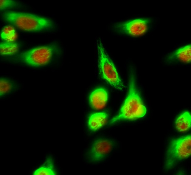
- Immunofluorescence analysis of Hela cell. 1,c-Fos Polyclonal Antibody(red) was diluted at 1:200(4° overnight). α-SMA Monoclonal Antibody(6A12)(green) was diluted at 1:200(4° overnight). 2, Goat Anti Rabbit Alexa Fluor 594 Catalog:RS3611 was diluted at 1:1000(room temperature, 50min). Goat Anti Mouse Alexa Fluor 488 Catalog:RS3208 was diluted at 1:1000(room temperature, 50min).
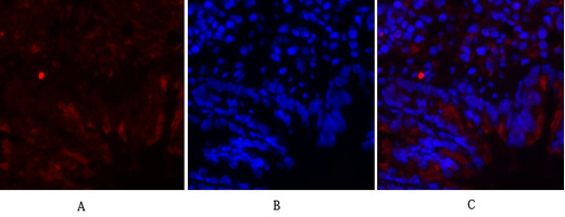
- Immunofluorescence analysis of rat-lung tissue. 1,c-Fos Polyclonal Antibody(red) was diluted at 1:200(4°C,overnight). 2, Cy3 labled Secondary antibody was diluted at 1:300(room temperature, 50min).3, Picture B: DAPI(blue) 10min. Picture A:Target. Picture B: DAPI. Picture C: merge of A+B
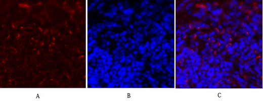
- Immunofluorescence analysis of rat-spleen tissue. 1,c-Fos Polyclonal Antibody(red) was diluted at 1:200(4°C,overnight). 2, Cy3 labled Secondary antibody was diluted at 1:300(room temperature, 50min).3, Picture B: DAPI(blue) 10min. Picture A:Target. Picture B: DAPI. Picture C: merge of A+B
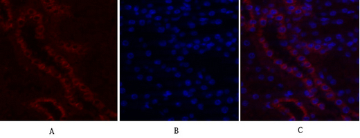
- Immunofluorescence analysis of mouse-kidney tissue. 1,c-Fos Polyclonal Antibody(red) was diluted at 1:200(4°C,overnight). 2, Cy3 labled Secondary antibody was diluted at 1:300(room temperature, 50min).3, Picture B: DAPI(blue) 10min. Picture A:Target. Picture B: DAPI. Picture C: merge of A+B
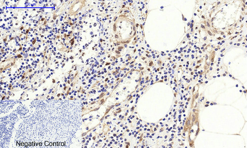
- Immunohistochemical analysis of paraffin-embedded Human-Appendix tissue. 1,c-Fos Polyclonal Antibody was diluted at 1:200(4°C,overnight). 2, Sodium citrate pH 6.0 was used for antibody retrieval(>98°C,20min). 3,Secondary antibody was diluted at 1:200(room tempeRature, 30min). Negative control was used by secondary antibody only.
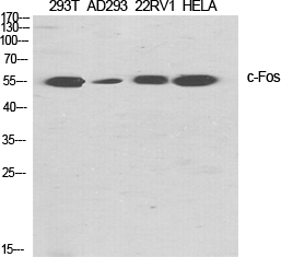
- Western Blot analysis of various cells using c-Fos Polyclonal Antibody diluted at 1:2000 cells nucleus extracted by Minute TM Cytoplasmic and Nuclear Fractionation kit (SC-003,Inventbiotech,MN,USA).


