AP-2γ Polyclonal Antibody
- Catalog No.:YT0254
- Applications:WB;IHC;IF;ELISA
- Reactivity:Human;Mouse;Rat
- Target:
- AP-2γ
- Gene Name:
- TFAP2C
- Protein Name:
- Transcription factor AP-2 gamma
- Human Gene Id:
- 7022
- Human Swiss Prot No:
- Q92754
- Mouse Gene Id:
- 21420
- Mouse Swiss Prot No:
- Q61312
- Immunogen:
- The antiserum was produced against synthesized peptide derived from human AP2C. AA range:401-450
- Specificity:
- AP-2γ Polyclonal Antibody detects endogenous levels of AP-2γ protein.
- Formulation:
- Liquid in PBS containing 50% glycerol, 0.5% BSA and 0.02% sodium azide.
- Source:
- Polyclonal, Rabbit,IgG
- Dilution:
- WB 1:500 - 1:2000. IHC 1:100 - 1:300. ELISA: 1:40000.. IF 1:50-200
- Purification:
- The antibody was affinity-purified from rabbit antiserum by affinity-chromatography using epitope-specific immunogen.
- Concentration:
- 1 mg/ml
- Storage Stability:
- -15°C to -25°C/1 year(Do not lower than -25°C)
- Other Name:
- TFAP2C;Transcription factor AP-2 gamma;AP2-gamma;Activating enhancer-binding protein 2 gamma;Transcription factor ERF-1
- Observed Band(KD):
- 45kD
- Background:
- transcription factor AP-2 gamma(TFAP2C) Homo sapiens The protein encoded by this gene is a sequence-specific DNA-binding transcription factor involved in the activation of several developmental genes. The encoded protein can act as either a homodimer or heterodimer with other family members and is induced during retinoic acid-mediated differentiation. It plays a role in the development of the eyes, face, body wall, limbs, and neural tube. [provided by RefSeq, Jul 2008],
- Function:
- domain:The WW-binding motif mediates interaction with WWOX.,function:Sequence-specific DNA-binding protein that interacts with inducible viral and cellular enhancer elements to regulate transcription of selected genes. AP-2 factors bind to the consensus sequence 5'-GCCNNNGGC-3' and activate genes involved in a large spectrum of important biological functions including proper eye, face, body wall, limb and neural tube development. They also suppress a number of genes including MCAM/MUC18, C/EBP alpha and MYC.,induction:During retinoic acid-mediated differentiation.,online information:Activatin protein 2 entry,PTM:Sumoylated on Lys-10; which inhibits transcriptional activity.,similarity:Belongs to the AP-2 family.,subunit:Binds DNA as a dimer. Can form homodimers or heterodimers with other AP-2 family members (By similarity). Interacts with WWOX. Interacts with CITED4. Interacts with UBE2I
- Subcellular Location:
- Nucleus .
- Expression:
- Liver,Mammary tumor,Ovary,Skin,
- June 19-2018
- WESTERN IMMUNOBLOTTING PROTOCOL
- June 19-2018
- IMMUNOHISTOCHEMISTRY-PARAFFIN PROTOCOL
- June 19-2018
- IMMUNOFLUORESCENCE PROTOCOL
- September 08-2020
- FLOW-CYTOMEYRT-PROTOCOL
- May 20-2022
- Cell-Based ELISA│解您多样本WB检测之困扰
- July 13-2018
- CELL-BASED-ELISA-PROTOCOL-FOR-ACETYL-PROTEIN
- July 13-2018
- CELL-BASED-ELISA-PROTOCOL-FOR-PHOSPHO-PROTEIN
- July 13-2018
- Antibody-FAQs
- Products Images
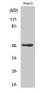
- Western Blot analysis of various cells using AP-2γ Polyclonal Antibody cells nucleus extracted by Minute TM Cytoplasmic and Nuclear Fractionation kit (SC-003,Inventbiotech,MN,USA).
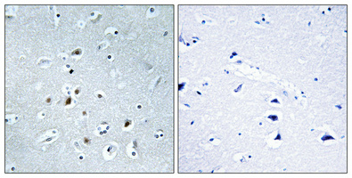
- Immunohistochemical analysis of paraffin-embedded Human brain. Antibody was diluted at 1:100(4° overnight). High-pressure and temperature Tris-EDTA,pH8.0 was used for antigen retrieval. Negetive contrl (right) obtaned from antibody was pre-absorbed by immunogen peptide.
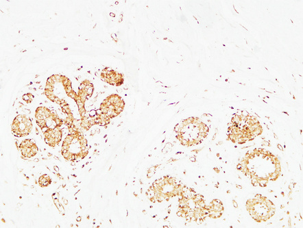
- Immunohistochemical analysis of paraffin-embedded Human breast. 1, Antibody was diluted at 1:200(4° overnight). 2, High-pressure and temperature EDTA, pH8.0 was used for antigen retrieval. 3,Secondary antibody was diluted at 1:200(room temperature, 30min).
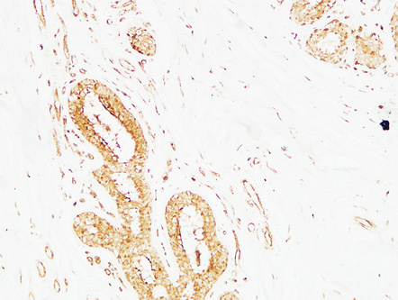
- Immunohistochemical analysis of paraffin-embedded Human breast. 1, Antibody was diluted at 1:200(4° overnight). 2, High-pressure and temperature EDTA, pH8.0 was used for antigen retrieval. 3,Secondary antibody was diluted at 1:200(room temperature, 30min).
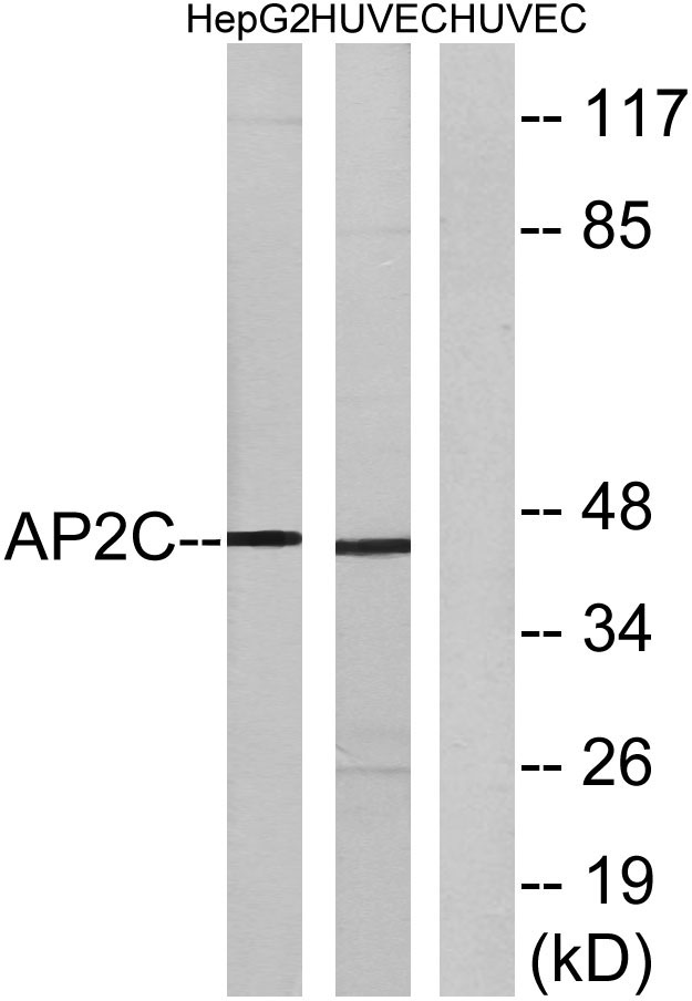
- Western blot analysis of lysates from HepG2 and HUVEC cells, using AP2C Antibody. The lane on the right is blocked with the synthesized peptide.

- Western blot analysis of the lysates from K562 cells using AP2C antibody.


