p38 (phospho Tyr323) Polyclonal Antibody
- Catalog No.:YP0718
- Applications:IF;WB;IHC;ELISA
- Reactivity:Human;Mouse;Rat
- Target:
- p38
- Fields:
- >>Endocrine resistance;>>MAPK signaling pathway;>>Rap1 signaling pathway;>>FoxO signaling pathway;>>Sphingolipid signaling pathway;>>Oocyte meiosis;>>Cellular senescence;>>Adrenergic signaling in cardiomyocytes;>>VEGF signaling pathway;>>Osteoclast differentiation;>>Signaling pathways regulating pluripotency of stem cells;>>Platelet activation;>>Neutrophil extracellular trap formation;>>Toll-like receptor signaling pathway;>>NOD-like receptor signaling pathway;>>RIG-I-like receptor signaling pathway;>>C-type lectin receptor signaling pathway;>>IL-17 signaling pathway;>>Th1 and Th2 cell differentiation;>>Th17 cell differentiation;>>T cell receptor signaling pathway;>>Fc epsilon RI signaling pathway;>>TNF signaling pathway;>>Leukocyte transendothelial migration;>>Thermogenesis;>>Neurotrophin signaling pathway;>>Retrograde endocannabinoid signaling;>>Dopaminergic synapse;>>Inflammatory mediator regulation of TRP channels;>>GnRH signaling pathway;>>Progesterone-mediated oocyte maturation;>
- Gene Name:
- MAPK14
- Protein Name:
- Mitogen-activated protein kinase 14
- Human Gene Id:
- 1432
- Human Swiss Prot No:
- Q16539
- Mouse Gene Id:
- 26416
- Mouse Swiss Prot No:
- P47811
- Rat Swiss Prot No:
- P70618
- Immunogen:
- The antiserum was produced against synthesized peptide derived from human p38 MAPK around the phosphorylation site of Tyr322. AA range:288-337
- Specificity:
- Phospho-p38 (Y323) Polyclonal Antibody detects endogenous levels of p38 protein only when phosphorylated at Y323.
- Formulation:
- Liquid in PBS containing 50% glycerol, 0.5% BSA and 0.02% sodium azide.
- Source:
- Polyclonal, Rabbit,IgG
- Dilution:
- IF 1:50-200 WB 1:500 - 1:2000. IHC 1:100 - 1:300. ELISA: 1:5000. Not yet tested in other applications.
- Purification:
- The antibody was affinity-purified from rabbit antiserum by affinity-chromatography using epitope-specific immunogen.
- Concentration:
- 1 mg/ml
- Storage Stability:
- -15°C to -25°C/1 year(Do not lower than -25°C)
- Other Name:
- MAPK14;CSBP;CSBP1;CSBP2;CSPB1;MXI2;SAPK2A;Mitogen-activated protein kinase 14;MAP kinase 14;MAPK 14;Cytokine suppressive anti-inflammatory drug-binding protein;CSAID-binding protein;CSBP;MAP kinase MXI2;MAX-interacting protein
- Observed Band(KD):
- 38kD
- Background:
- The protein encoded by this gene is a member of the MAP kinase family. MAP kinases act as an integration point for multiple biochemical signals, and are involved in a wide variety of cellular processes such as proliferation, differentiation, transcription regulation and development. This kinase is activated by various environmental stresses and proinflammatory cytokines. The activation requires its phosphorylation by MAP kinase kinases (MKKs), or its autophosphorylation triggered by the interaction of MAP3K7IP1/TAB1 protein with this kinase. The substrates of this kinase include transcription regulator ATF2, MEF2C, and MAX, cell cycle regulator CDC25B, and tumor suppressor p53, which suggest the roles of this kinase in stress related transcription and cell cycle regulation, as well as in genotoxic stress response. Four alternatively spliced transcript variants of this gene encoding d
- Function:
- catalytic activity:ATP + a protein = ADP + a phosphoprotein.,cofactor:Magnesium.,domain:The TXY motif contains the threonine and tyrosine residues whose phosphorylation activates the MAP kinases.,enzyme regulation:Activated by threonine and tyrosine phosphorylation by either of two dual specificity kinases, MAP2K3 or MAP2K6, and potentially also MAP2K4. Inhibited by dual specificity phosphatases, such as DUSP1. Specifically inhibited by the binding of pyridinyl-imidazole compounds, which are cytokine-suppressive anti-inflammatory drugs (CSAID). Isoform Mxi2 is 100-fold less sensitive to these agents than the other isoforms and is not inhibited by DUSP1. Isoform Exip is not activated by MAP2K6.,function:Responds to activation by environmental stress, pro-inflammatory cytokines and lipopolysaccharide (LPS) by phosphorylating a number of transcription factors, such as ELK1 and ATF2 and seve
- Subcellular Location:
- Cytoplasm . Nucleus .
- Expression:
- Brain, heart, placenta, pancreas and skeletal muscle. Expressed to a lesser extent in lung, liver and kidney.
Interleukin-33 Promotes Cell Survival via p38 MAPK-Mediated Interleukin-6 Gene Expression and Release in Pediatric AML. Frontiers in Immunology Front Immunol. 2020; 11: 595053 WB Human bone marrow (BM),peripheral blood (PB) HL-60 cell
- June 19-2018
- WESTERN IMMUNOBLOTTING PROTOCOL
- June 19-2018
- IMMUNOHISTOCHEMISTRY-PARAFFIN PROTOCOL
- June 19-2018
- IMMUNOFLUORESCENCE PROTOCOL
- September 08-2020
- FLOW-CYTOMEYRT-PROTOCOL
- May 20-2022
- Cell-Based ELISA│解您多样本WB检测之困扰
- July 13-2018
- CELL-BASED-ELISA-PROTOCOL-FOR-ACETYL-PROTEIN
- July 13-2018
- CELL-BASED-ELISA-PROTOCOL-FOR-PHOSPHO-PROTEIN
- July 13-2018
- Antibody-FAQs
- Products Images
if-human-stomach129.jpg)
- Immunofluorescence analysis of human-stomach tissue. 1,p38 (phospho Tyr323) Polyclonal Antibody(red) was diluted at 1:200(4°C,overnight). 2, Cy3 labled Secondary antibody was diluted at 1:300(room temperature, 50min).3, Picture B: DAPI(blue) 10min. Picture A:Target. Picture B: DAPI. Picture C: merge of A+B
if-human-stomach130.jpg)
- Immunofluorescence analysis of human-stomach tissue. 1,p38 (phospho Tyr323) Polyclonal Antibody(red) was diluted at 1:200(4°C,overnight). 2, Cy3 labled Secondary antibody was diluted at 1:300(room temperature, 50min).3, Picture B: DAPI(blue) 10min. Picture A:Target. Picture B: DAPI. Picture C: merge of A+B
poly-ihc-human-uterus.jpg)
- Immunohistochemical analysis of paraffin-embedded Human-uterus tissue. 1,p38 (phospho Tyr323) Polyclonal Antibody was diluted at 1:200(4°C,overnight). 2, Sodium citrate pH 6.0 was used for antibody retrieval(>98°C,20min). 3,Secondary antibody was diluted at 1:200(room tempeRature, 30min). Negative control was used by secondary antibody only.
poly-ihc-human-lung.jpg)
- Immunohistochemical analysis of paraffin-embedded Human-lung tissue. 1,p38 (phospho Tyr323) Polyclonal Antibody was diluted at 1:200(4°C,overnight). 2, Sodium citrate pH 6.0 was used for antibody retrieval(>98°C,20min). 3,Secondary antibody was diluted at 1:200(room tempeRature, 30min). Negative control was used by secondary antibody only.
poly-ihc-rat-heart.jpg)
- Immunohistochemical analysis of paraffin-embedded Rat-heart tissue. 1,p38 (phospho Tyr323) Polyclonal Antibody was diluted at 1:200(4°C,overnight). 2, Sodium citrate pH 6.0 was used for antibody retrieval(>98°C,20min). 3,Secondary antibody was diluted at 1:200(room tempeRature, 30min). Negative control was used by secondary antibody only.
poly-ihc-rat-lung.jpg)
- Immunohistochemical analysis of paraffin-embedded Rat-lung tissue. 1,p38 (phospho Tyr323) Polyclonal Antibody was diluted at 1:200(4°C,overnight). 2, Sodium citrate pH 6.0 was used for antibody retrieval(>98°C,20min). 3,Secondary antibody was diluted at 1:200(room tempeRature, 30min). Negative control was used by secondary antibody only.
poly-ihc-rat-kidney.jpg)
- Immunohistochemical analysis of paraffin-embedded Rat-kidney tissue. 1,p38 (phospho Tyr323) Polyclonal Antibody was diluted at 1:200(4°C,overnight). 2, Sodium citrate pH 6.0 was used for antibody retrieval(>98°C,20min). 3,Secondary antibody was diluted at 1:200(room tempeRature, 30min). Negative control was used by secondary antibody only.
poly-ihc-rat-spleen.jpg)
- Immunohistochemical analysis of paraffin-embedded Rat-spleen tissue. 1,p38 (phospho Tyr323) Polyclonal Antibody was diluted at 1:200(4°C,overnight). 2, Sodium citrate pH 6.0 was used for antibody retrieval(>98°C,20min). 3,Secondary antibody was diluted at 1:200(room tempeRature, 30min). Negative control was used by secondary antibody only.
poly-ihc-mouse-lung.jpg)
- Immunohistochemical analysis of paraffin-embedded Mouse-lung tissue. 1,p38 (phospho Tyr323) Polyclonal Antibody was diluted at 1:200(4°C,overnight). 2, Sodium citrate pH 6.0 was used for antibody retrieval(>98°C,20min). 3,Secondary antibody was diluted at 1:200(room tempeRature, 30min). Negative control was used by secondary antibody only.
poly-ihc-mouse-kidney.jpg)
- Immunohistochemical analysis of paraffin-embedded Mouse-kidney tissue. 1,p38 (phospho Tyr323) Polyclonal Antibody was diluted at 1:200(4°C,overnight). 2, Sodium citrate pH 6.0 was used for antibody retrieval(>98°C,20min). 3,Secondary antibody was diluted at 1:200(room tempeRature, 30min). Negative control was used by secondary antibody only.

- Western Blot analysis of various cells using Phospho-p38 (Y323) Polyclonal Antibody diluted at 1:500

- The picture was kindly provided by our customer.

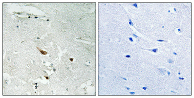
- Immunohistochemical analysis of paraffin-embedded Human brain. Antibody was diluted at 1:100(4° overnight). High-pressure and temperature Tris-EDTA,pH8.0 was used for antigen retrieval. Negetive contrl (right) obtaned from antibody was pre-absorbed by immunogen peptide.

- Enzyme-Linked Immunosorbent Assay (Phospho-ELISA) for Immunogen Phosphopeptide (Phospho-left) and Non-Phosphopeptide (Phospho-right), using p38 MAPK (Phospho-Tyr322) Antibody

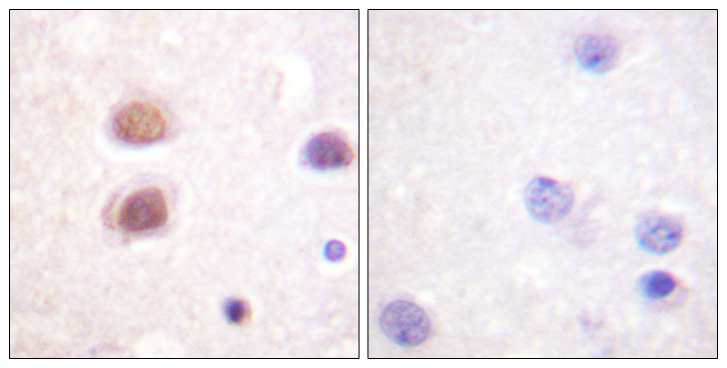
- Immunohistochemistry analysis of paraffin-embedded human brain, using p38 MAPK (Phospho-Tyr322) Antibody. The picture on the right is blocked with the phospho peptide.

- Western blot analysis of lysates from Jurkat cells, using p38 MAPK (Phospho-Tyr322) Antibody. The lane on the right is blocked with the phospho peptide.


