CD15 Monoclonal Antibody(Q89)
- Catalog No.:YM3105
- Applications:IHC;IF
- Reactivity:Human
- Target:
- CD15
- Fields:
- >>Mannose type O-glycan biosynthesis;>>Glycosphingolipid biosynthesis - lacto and neolacto series;>>Metabolic pathways
- Gene Name:
- FUT4
- Protein Name:
- Alpha-(1,3)-fucosyltransferase
- Human Gene Id:
- 2526
- Human Swiss Prot No:
- P22083
- Mouse Gene Id:
- 14345
- Mouse Swiss Prot No:
- Q11127
- Rat Swiss Prot No:
- Q62994
- Immunogen:
- Synthetic Peptide of CD15
- Specificity:
- The antibody detects endogenous CD15 protein.
- Formulation:
- PBS, pH 7.4, containing 0.5%BSA, 0.02% sodium azide as Preservative and 50% Glycerol.
- Source:
- Monoclonal, Mouse
- Dilution:
- IHC 1:200 IF 1:50-200
- Purification:
- The antibody was affinity-purified from mouse ascites by affinity-chromatography using specific immunogen.
- Storage Stability:
- -15°C to -25°C/1 year(Do not lower than -25°C)
- Other Name:
- FUT4;ELFT;FCT3A;Alpha-(1,3)-fucosyltransferase;ELAM-1 ligand fucosyltransferase;Fucosyltransferase 4;Fucosyltransferase IV;Fuc-TIV;FucT-IV;Galactoside 3-L-fucosyltransferase
- Molecular Weight(Da):
- 46kD
- Background:
- The product of this gene transfers fucose to N-acetyllactosamine polysaccharides to generate fucosylated carbohydrate structures. It catalyzes the synthesis of the non-sialylated antigen, Lewis x (CD15). [provided by RefSeq, Jan 2009],
- Function:
- caution:It is uncertain whether Met-1 or Met-126 is the initiator.,function:May catalyze alpha-1,3 glycosidic linkages involved in the expression of Lewis X/SSEA-1 and VIM-2 antigens.,online information:Fucosyltransferase 4,online information:GlycoGene database,pathway:Protein modification; protein glycosylation.,similarity:Belongs to the glycosyltransferase 10 family.,subcellular location:Membrane-bound form in trans cisternae of Golgi.,
- Subcellular Location:
- Golgi apparatus, Golgi stack membrane; Single-pass type II membrane protein. Membrane-bound form in trans cisternae of Golgi.
- Expression:
- [Isoform Short]: Expressed at low levels in bone marrow-derived mesenchymal stem cells. ; Expressed in cord blood immature promyelocytes and in peripheral blood myeloid and lymphoid cell populations.
- June 19-2018
- WESTERN IMMUNOBLOTTING PROTOCOL
- June 19-2018
- IMMUNOHISTOCHEMISTRY-PARAFFIN PROTOCOL
- June 19-2018
- IMMUNOFLUORESCENCE PROTOCOL
- September 08-2020
- FLOW-CYTOMEYRT-PROTOCOL
- May 20-2022
- Cell-Based ELISA│解您多样本WB检测之困扰
- July 13-2018
- CELL-BASED-ELISA-PROTOCOL-FOR-ACETYL-PROTEIN
- July 13-2018
- CELL-BASED-ELISA-PROTOCOL-FOR-PHOSPHO-PROTEIN
- July 13-2018
- Antibody-FAQs
- Products Images

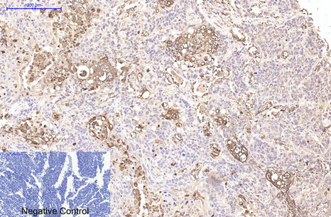
- Immunohistochemical analysis of paraffin-embedded Human-lung-cancer tissue. 1,CD15 Monoclonal Antibody(Q89) was diluted at 1:200(4°C,overnight). 2, Sodium citrate pH 6.0 was used for antibody retrieval(>98°C,20min). 3,Secondary antibody was diluted at 1:200(room tempeRature, 30min). Negative control was used by secondary antibody only.

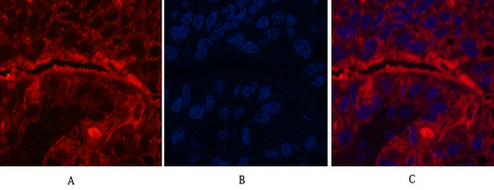
- Immunofluorescence analysis of Human-liver-cancer tissue. 1,CD15 Monoclonal Antibody(Q89)(red) was diluted at 1:200(4°C,overnight). 2, Cy3 labled Secondary antibody was diluted at 1:300(room temperature, 50min).3, Picture B: DAPI(blue) 10min. Picture A:Target. Picture B: DAPI. Picture C: merge of A+B

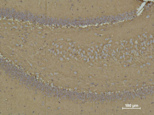
- Immunohistochemical analysis of paraffin-embedded Rat Brain Tissue using CD 15 Mouse mAb diluted at 1:500.

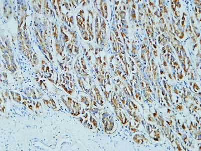
- Immunohistochemical analysis of paraffin-embedded Human stomach. 1, Antibody was diluted at 1:200(4° overnight). 2, High-pressure and temperature EDTA, pH8.0 was used for antigen retrieval. 3,Secondary antibody was diluted at 1:200(room temperature, 30min).


