CD21 Monoclonal Antibody(2C5)
- Catalog No.:YM3060
- Applications:IHC;IF
- Reactivity:Human;Mouse;Rat
- Target:
- CD21
- Fields:
- >>Complement and coagulation cascades;>>Hematopoietic cell lineage;>>B cell receptor signaling pathway;>>Epstein-Barr virus infection
- Gene Name:
- CR2
- Protein Name:
- Complement receptor type 2
- Human Gene Id:
- 1380
- Human Swiss Prot No:
- P20023
- Mouse Swiss Prot No:
- P19070
- Immunogen:
- Synthetic Peptide of CD21
- Specificity:
- The antibody detects endogenous CD21 proteins.
- Formulation:
- PBS, pH 7.4, containing 0.5%BSA, 0.02% sodium azide as Preservative and 50% Glycerol.
- Source:
- Monoclonal, Mouse
- Dilution:
- IHC 1:200 IF 1:50-200
- Purification:
- The antibody was affinity-purified from mouse ascites by affinity-chromatography using specific immunogen.
- Storage Stability:
- -15°C to -25°C/1 year(Do not lower than -25°C)
- Other Name:
- CR2;C3DR;Complement receptor type 2;Cr2;Complement C3d receptor;Epstein-Barr virus receptor;EBV receptor;CD21
- Observed Band(KD):
- 113kD
- Background:
- This gene encodes a membrane protein, which functions as a receptor for Epstein-Barr virus (EBV) binding on B and T lymphocytes. Genetic variations in this gene are associated with susceptibility to systemic lupus erythematosus type 9 (SLEB9). Alternatively spliced transcript variants encoding different isoforms have been found for this gene.[provided by RefSeq, Sep 2009],
- Function:
- disease:Genetic variations in CR2 are associated with susceptibility to systemic lupus erythematosus type 9 (SLEB9) [MIM:610927]. Systemic lupus erythematosus (SLE) is a chronic autoimmune disease with a complex genetic basis. SLE is an inflammatory, and often febrile multisystemic disorder of connective tissue characterized principally by involvement of the skin, joints, kidneys, and serosal membranes. It is thought to represent a failure of the regulatory mechanisms of the autoimmune system.,function:Receptor for complement C3Dd, for the Epstein-Barr virus on human B-cells and T-cells and for HNRPU. Participates in B lymphocytes activation.,similarity:Belongs to the receptors of complement activation (RCA) family.,similarity:Contains 15 Sushi (CCP/SCR) domains.,tissue specificity:Mature B-lymphocytes, T-lymphocytes, pharyngeal epithelial cells, astrocytes and follicular dendritic cells
- Subcellular Location:
- Cell membrane ; Single-pass type I membrane protein.
- Expression:
- Mature B-lymphocytes, T-lymphocytes, pharyngeal epithelial cells, astrocytes and follicular dendritic cells of the spleen.
- June 19-2018
- WESTERN IMMUNOBLOTTING PROTOCOL
- June 19-2018
- IMMUNOHISTOCHEMISTRY-PARAFFIN PROTOCOL
- June 19-2018
- IMMUNOFLUORESCENCE PROTOCOL
- September 08-2020
- FLOW-CYTOMEYRT-PROTOCOL
- May 20-2022
- Cell-Based ELISA│解您多样本WB检测之困扰
- July 13-2018
- CELL-BASED-ELISA-PROTOCOL-FOR-ACETYL-PROTEIN
- July 13-2018
- CELL-BASED-ELISA-PROTOCOL-FOR-PHOSPHO-PROTEIN
- July 13-2018
- Antibody-FAQs
- Products Images
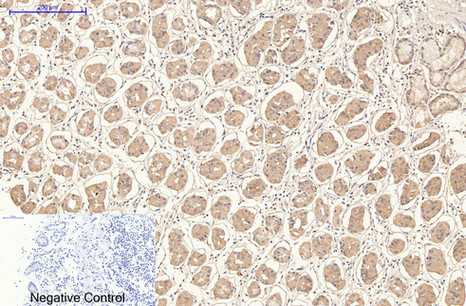
- Immunohistochemical analysis of paraffin-embedded Human-stomach tissue. 1,CD21 Monoclonal Antibody(2C5) was diluted at 1:200(4°C,overnight). 2, Sodium citrate pH 6.0 was used for antibody retrieval(>98°C,20min). 3,Secondary antibody was diluted at 1:200(room tempeRature, 30min). Negative control was used by secondary antibody only.
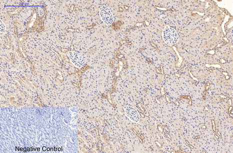
- Immunohistochemical analysis of paraffin-embedded Rat-kidney tissue. 1,CD21 Monoclonal Antibody(2C5) was diluted at 1:200(4°C,overnight). 2, Sodium citrate pH 6.0 was used for antibody retrieval(>98°C,20min). 3,Secondary antibody was diluted at 1:200(room tempeRature, 30min). Negative control was used by secondary antibody only.
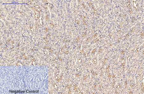
- Immunohistochemical analysis of paraffin-embedded Mouse-kidney tissue. 1,CD21 Monoclonal Antibody(2C5) was diluted at 1:200(4°C,overnight). 2, Sodium citrate pH 6.0 was used for antibody retrieval(>98°C,20min). 3,Secondary antibody was diluted at 1:200(room tempeRature, 30min). Negative control was used by secondary antibody only.
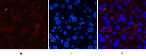
- Immunofluorescence analysis of Mouse-liver tissue. 1,CD21 Monoclonal Antibody(2C5)(red) was diluted at 1:200(4°C,overnight). 2, Cy3 labled Secondary antibody was diluted at 1:300(room temperature, 50min).3, Picture B: DAPI(blue) 10min. Picture A:Target. Picture B: DAPI. Picture C: merge of A+B
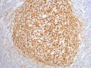
- IHC staining of human tonsil tissue, diluted at 1:200.


