Rab 25 Monoclonal Antibody
- Catalog No.:YM0546
- Applications:WB;IHC;IF;FCM;ELISA
- Reactivity:Human;Mouse
- Target:
- Rab 25
- Gene Name:
- RAB25
- Protein Name:
- Ras-related protein Rab-25
- Human Gene Id:
- 57111
- Human Swiss Prot No:
- P57735
- Mouse Gene Id:
- 53868
- Mouse Swiss Prot No:
- Q9WTL2
- Immunogen:
- Purified recombinant fragment of Rab 25 expressed in E. Coli.
- Specificity:
- Rab 25 Monoclonal Antibody detects endogenous levels of Rab 25 protein.
- Formulation:
- Liquid in PBS containing 50% glycerol, 0.5% BSA and 0.02% sodium azide.
- Source:
- Monoclonal, Mouse
- Dilution:
- WB 1:500 - 1:2000. IHC 1:200 - 1:1000. IF 1:200 - 1:1000. Flow cytometry: 1:200 - 1:400. ELISA: 1:10000. Not yet tested in other applications.
- Purification:
- Affinity purification
- Storage Stability:
- -15°C to -25°C/1 year(Do not lower than -25°C)
- Other Name:
- RAB25;CATX8;Ras-related protein Rab-25;CATX-8
- Molecular Weight(Da):
- 23kD
- References:
- 1. JR Goldenring, KR Shen, HD Vaughan. et al. J. Biol. Chem,1993,268(25):18419-18422.
2. Xiaoye W, Ravindra K, Jennifer N. et al. J. Biol. Chem,2000,275(37):29138-29146.
- Background:
- The protein encoded by this gene is a member of the RAS superfamily of small GTPases. The encoded protein is involved in membrane trafficking and cell survival. This gene has been found to be a tumor suppressor and an oncogene, depending on the context. Two variants, one protein-coding and the other not, have been found for this gene. [provided by RefSeq, Nov 2015],
- Function:
- function:May selectively regulate the apical recycling and/or transcytotic pathways.,similarity:Belongs to the small GTPase superfamily. Rab family.,subunit:Interacts with RAB11FIP1, RAB11FIP2, RAB11FIP3 and RAB11FIP4.,
- Subcellular Location:
- Cell membrane ; Lipid-anchor ; Cytoplasmic side . Cell projection, pseudopodium membrane . Cytoplasmic vesicle . Colocalizes with integrin alpha-V/beta-1 in vesicles at the pseudopodial tips. .
- Expression:
- Expressed in ovarian epithelium (NOE) and breast tissue. Expressed in ovarian cancer; expression is increased relative to NOE cells. Expression in ovarian cancer is stage dependent, with stage III and stage IV showing higher levels than early stage cancers. Expressed in breast cancer; expression is increased relative to normal breast tissue.
- June 19-2018
- WESTERN IMMUNOBLOTTING PROTOCOL
- June 19-2018
- IMMUNOHISTOCHEMISTRY-PARAFFIN PROTOCOL
- June 19-2018
- IMMUNOFLUORESCENCE PROTOCOL
- September 08-2020
- FLOW-CYTOMEYRT-PROTOCOL
- May 20-2022
- Cell-Based ELISA│解您多样本WB检测之困扰
- July 13-2018
- CELL-BASED-ELISA-PROTOCOL-FOR-ACETYL-PROTEIN
- July 13-2018
- CELL-BASED-ELISA-PROTOCOL-FOR-PHOSPHO-PROTEIN
- July 13-2018
- Antibody-FAQs
- Products Images

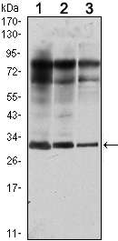
- Western Blot analysis using Rab 25 Monoclonal Antibody against MCF-7 (1), T47D (2) and GC7901 (3) cell lysate.

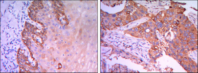
- Immunohistochemistry analysis of paraffin-embedded esophagus tissues (left) and human lung cancer (right) with DAB staining using Rab 25 Monoclonal Antibody.

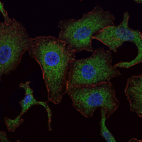
- Immunofluorescence analysis of A549 cells using Rab 25 Monoclonal Antibody (green). Blue: DRAQ5 fluorescent DNA dye. Red: Actin filaments have been labeled with Alexa Fluor-555 phalloidin.

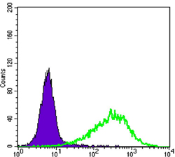
- Flow cytometric analysis of NIH/3T3 cells using Rab 25 Monoclonal Antibody (green) and negative control (purple).


