PD-1 rabbit pAb
- Catalog No.:YT7834
- Applications:WB;ELISA;IHC
- Reactivity:Human;Mouse
- Target:
- PD1
- Fields:
- >>Cell adhesion molecules;>>T cell receptor signaling pathway;>>PD-L1 expression and PD-1 checkpoint pathway in cancer
- Gene Name:
- PDCD1 PD1
- Protein Name:
- Pdcd-1
- Human Gene Id:
- 5133
- Human Swiss Prot No:
- Q15116
- Mouse Gene Id:
- 18566
- Mouse Swiss Prot No:
- Q02242
- Immunogen:
- Synthesized peptide derived from human Pdcd-1 AA range: 80-130
- Specificity:
- This antibody detects endogenous levels of Human Pdcd-1
- Formulation:
- Liquid in PBS containing 50% glycerol, 0.5% BSA and 0.02% sodium azide.
- Source:
- Polyclonal, Rabbit,IgG
- Dilution:
- WB 1:500-2000;IHC 1:50-300; ELISA 2000-20000
- Purification:
- The antibody was affinity-purified from rabbit antiserum by affinity-chromatography using epitope-specific immunogen.
- Concentration:
- 1 mg/ml
- Storage Stability:
- -15°C to -25°C/1 year(Do not lower than -25°C)
- Other Name:
- Programmed cell death protein 1 (Protein PD-1;hPD-1;CD antigen CD279)
- Molecular Weight(Da):
- 32kD
- Background:
- This gene encodes a cell surface membrane protein of the immunoglobulin superfamily. This protein is expressed in pro-B-cells and is thought to play a role in their differentiation. In mice, expression of this gene is induced in the thymus when anti-CD3 antibodies are injected and large numbers of thymocytes undergo apoptosis. Mice deficient for this gene bred on a BALB/c background developed dilated cardiomyopathy and died from congestive heart failure. These studies suggest that this gene product may also be important in T cell function and contribute to the prevention of autoimmune diseases. [provided by RefSeq, Jul 2008],
- Function:
- developmental stage:Induced at programmed cell death.,disease:Genetic variation in PDCD1 is associated with susceptibility to systemic lupus erythematosus type 2 (SLEB2) [MIM:605218]. Systemic lupus erythematosus is a chronic, inflammatory and often febrile multisystemic disorder of connective tissue. It affects principally the skin, joints, kidneys and serosal membranes. It is thought to represent a failure of the regulatory mechanisms of the autoimmune system.,function:Possible cell death inducer, in association with other factors.,similarity:Contains 1 Ig-like V-type (immunoglobulin-like) domain.,subunit:Monomer.,
- Subcellular Location:
- Cell membrane ; Single-pass type I membrane protein.
- June 19-2018
- WESTERN IMMUNOBLOTTING PROTOCOL
- June 19-2018
- IMMUNOHISTOCHEMISTRY-PARAFFIN PROTOCOL
- June 19-2018
- IMMUNOFLUORESCENCE PROTOCOL
- September 08-2020
- FLOW-CYTOMEYRT-PROTOCOL
- May 20-2022
- Cell-Based ELISA│解您多样本WB检测之困扰
- July 13-2018
- CELL-BASED-ELISA-PROTOCOL-FOR-ACETYL-PROTEIN
- July 13-2018
- CELL-BASED-ELISA-PROTOCOL-FOR-PHOSPHO-PROTEIN
- July 13-2018
- Antibody-FAQs
- Products Images
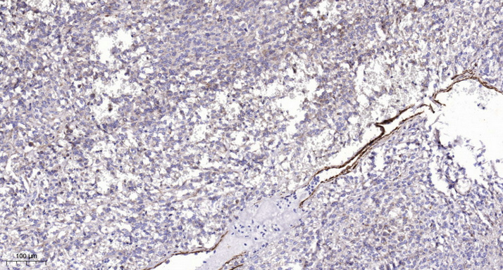
- Immunohistochemical analysis of paraffin-embedded human Colon cancer. 1, Antibody was diluted at 1:200(4° overnight). 2, Tris-EDTA,pH9.0 was used for antigen retrieval. 3,Secondary antibody was diluted at 1:200(room temperature, 45min).


