α-protein Kinase 1 Polyclonal Antibody
- Catalog No.:YT5001
- Applications:WB;IHC;IF;ELISA
- Reactivity:Human;Mouse
- Target:
- α-protein Kinase 1
- Gene Name:
- ALPK1
- Protein Name:
- Alpha-protein kinase 1
- Human Gene Id:
- 80216
- Human Swiss Prot No:
- Q96QP1
- Mouse Swiss Prot No:
- Q9CXB8
- Immunogen:
- The antiserum was produced against synthesized peptide derived from human ALPK1. AA range:11-60
- Specificity:
- α-protein Kinase 1 Polyclonal Antibody detects endogenous levels of α-protein Kinase 1 protein.
- Formulation:
- Liquid in PBS containing 50% glycerol, 0.5% BSA and 0.02% sodium azide.
- Source:
- Polyclonal, Rabbit,IgG
- Dilution:
- WB 1:500 - 1:2000. IHC 1:100 - 1:300. IF 1:200 - 1:1000. ELISA: 1:20000. Not yet tested in other applications.
- Purification:
- The antibody was affinity-purified from rabbit antiserum by affinity-chromatography using epitope-specific immunogen.
- Concentration:
- 1 mg/ml
- Storage Stability:
- -15°C to -25°C/1 year(Do not lower than -25°C)
- Other Name:
- ALPK1;KIAA1527;LAK;Alpha-protein kinase 1;Chromosome 4 kinase;Lymphocyte alpha-protein kinase
- Observed Band(KD):
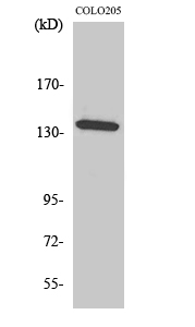
- 139kD
- Background:
- This gene encodes an alpha kinase. Mice which were homozygous for disrupted copies of this gene exhibited coordination defects (PMID: 21208416). Multiple transcript variants encoding different isoforms have been found for this gene. [provided by RefSeq, Dec 2011],
- Function:
- function:Kinases that recognize phosphorylation sites in which the surrounding peptides have an alpha-helical conformation.,similarity:Belongs to the protein kinase superfamily. Alpha-type protein kinase family. ALPK subfamily.,similarity:Contains 1 alpha-type protein kinase domain.,tissue specificity:Highly expressed in liver.,
- Subcellular Location:
- Cytoplasm, cytosol . Cytoplasm, cytoskeleton, spindle pole . Cytoplasm, cytoskeleton, microtubule organizing center, centrosome . Cell projection, cilium . Localized at the base of primary cilia. .
- Expression:
- Highly expressed in liver. Expressed in the optic nerve and retinal pigmented epithelium. Lower expression is observed in the macula and extramacular retina (PubMed:30967659).
Alpha‐kinase1 promotes tubular injury and interstitial inflammation in diabetic nephropathy by canonical pyroptosis pathway BIOLOGICAL RESEARCH Xuejing Zhu IHC,WB Human,Mouse Renal HK-2 cell
- June 19-2018
- WESTERN IMMUNOBLOTTING PROTOCOL
- June 19-2018
- IMMUNOHISTOCHEMISTRY-PARAFFIN PROTOCOL
- June 19-2018
- IMMUNOFLUORESCENCE PROTOCOL
- September 08-2020
- FLOW-CYTOMEYRT-PROTOCOL
- May 20-2022
- Cell-Based ELISA│解您多样本WB检测之困扰
- July 13-2018
- CELL-BASED-ELISA-PROTOCOL-FOR-ACETYL-PROTEIN
- July 13-2018
- CELL-BASED-ELISA-PROTOCOL-FOR-PHOSPHO-PROTEIN
- July 13-2018
- Antibody-FAQs
- Products Images

- Western Blot analysis of various cells using α-protein Kinase 1 Polyclonal Antibody. Secondary antibody(catalog#:RS0002) was diluted at 1:20000

- Western blot analysis of lysates from COLO and HepG2 cells, using ALPK1 Antibody. The lane on the right is blocked with the synthesized peptide.

- Western blot analysis of the lysates from 293 cells using ALPK1 antibody.

- Immunohistochemical analysis of paraffin-embedded human Gastric adenocarcinoma. 1, Antibody was diluted at 1:200(4° overnight). 2, Tris-EDTA,pH9.0 was used for antigen retrieval. 3,Secondary antibody was diluted at 1:200(room temperature, 45min).


