XPG Polyclonal Antibody
- Catalog No.:YT4915
- Applications:WB;IHC;IF;ELISA
- Reactivity:Human;Mouse
- Target:
- XPG
- Fields:
- >>Nucleotide excision repair
- Gene Name:
- ERCC5
- Protein Name:
- DNA repair protein complementing XP-G cells
- Human Gene Id:
- 2073
- Human Swiss Prot No:
- P28715
- Mouse Swiss Prot No:
- P35689
- Immunogen:
- The antiserum was produced against synthesized peptide derived from human ERCC5. AA range:131-180
- Specificity:
- XPG Polyclonal Antibody detects endogenous levels of XPG protein.
- Formulation:
- Liquid in PBS containing 50% glycerol, 0.5% BSA and 0.02% sodium azide.
- Source:
- Polyclonal, Rabbit,IgG
- Dilution:
- WB 1:500 - 1:2000. IHC 1:100 - 1:300. IF 1:200 - 1:1000. ELISA: 1:5000. Not yet tested in other applications.
- Purification:
- The antibody was affinity-purified from rabbit antiserum by affinity-chromatography using epitope-specific immunogen.
- Concentration:
- 1 mg/ml
- Storage Stability:
- -15°C to -25°C/1 year(Do not lower than -25°C)
- Other Name:
- ERCC5;ERCM2;XPG;XPGC;DNA repair protein complementing XP-G cells;DNA excision repair protein ERCC-5;Xeroderma pigmentosum group G-complementing protein
- Observed Band(KD):
- 130kD
- Background:
- This gene encodes a single-strand specific DNA endonuclease that makes the 3' incision in DNA excision repair following UV-induced damage. The protein may also function in other cellular processes, including RNA polymerase II transcription, and transcription-coupled DNA repair. Mutations in this gene cause xeroderma pigmentosum complementation group G (XP-G), which is also referred to as xeroderma pigmentosum VII (XP7), a skin disorder characterized by hypersensitivity to UV light and increased susceptibility for skin cancer development following UV exposure. Some patients also develop Cockayne syndrome, which is characterized by severe growth defects, mental retardation, and cachexia. Read-through transcription exists between this gene and the neighboring upstream BIVM (basic, immunoglobulin-like variable motif containing) gene. [provided by RefSeq, Feb 2011],
- Function:
- cofactor:Binds 2 magnesium ions per subunit. They probably participate in the reaction catalyzed by the enzyme. May bind an additional third magnesium ion after substrate binding.,disease:Defects in ERCC5 are the cause of xeroderma pigmentosum complementation group G (XP-G) [MIM:278780]; also known as xeroderma pigmentosum VII (XP7). Xeroderma pigmentosum is an autosomal recessive pigmentary skin disorder characterized by solar hypersensitivity of the skin, high predisposition for developing cancers on areas exposed to sunlight and, in some cases, neurological abnormalities. Some XP-G patients present features of Cockayne syndrome, including dwarfism, sensorineural deafness, microcephaly, mental retardation, pigmentary retinopathy, ataxia, decreased nerve conduction velocities.,function:Single-stranded structure-specific DNA endonuclease involved in DNA excision repair. Makes the 3'incis
- Subcellular Location:
- Nucleus . Chromosome . Colocalizes with RAD51 to nuclear foci in S phase (PubMed:26833090). Localizes to DNA double-strand breaks (DBS) during replication stress (PubMed:26833090). Colocalizes with BRCA2 to nuclear foci following DNA replication stress (PubMed:26833090). .
- Expression:
- Bone marrow,Epithelium,Eye,
- June 19-2018
- WESTERN IMMUNOBLOTTING PROTOCOL
- June 19-2018
- IMMUNOHISTOCHEMISTRY-PARAFFIN PROTOCOL
- June 19-2018
- IMMUNOFLUORESCENCE PROTOCOL
- September 08-2020
- FLOW-CYTOMEYRT-PROTOCOL
- May 20-2022
- Cell-Based ELISA│解您多样本WB检测之困扰
- July 13-2018
- CELL-BASED-ELISA-PROTOCOL-FOR-ACETYL-PROTEIN
- July 13-2018
- CELL-BASED-ELISA-PROTOCOL-FOR-PHOSPHO-PROTEIN
- July 13-2018
- Antibody-FAQs
- Products Images
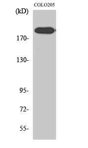
- Western Blot analysis of various cells using XPG Polyclonal Antibody diluted at 1:2000. Secondary antibody(catalog#:RS0002) was diluted at 1:20000 cells nucleus extracted by Minute TM Cytoplasmic and Nuclear Fractionation kit (SC-003,Inventbiotech,MN,USA).
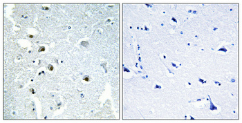
- Immunohistochemical analysis of paraffin-embedded Human brain. Antibody was diluted at 1:100(4° overnight). High-pressure and temperature Tris-EDTA,pH8.0 was used for antigen retrieval. Negetive contrl (right) obtaned from antibody was pre-absorbed by immunogen peptide.
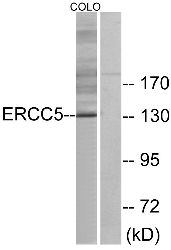
- Western blot analysis of lysates from COLO cells, using ERCC5 Antibody. The lane on the right is blocked with the synthesized peptide.


