XIAP Polyclonal Antibody
- Catalog No.:YT4913
- Applications:WB;IHC;IF;ELISA
- Reactivity:Human;Mouse;Rat
- Target:
- XIAP
- Fields:
- >>Platinum drug resistance;>>NF-kappa B signaling pathway;>>Ubiquitin mediated proteolysis;>>Apoptosis;>>Apoptosis - multiple species;>>Necroptosis;>>Focal adhesion;>>NOD-like receptor signaling pathway;>>Toxoplasmosis;>>Human T-cell leukemia virus 1 infection;>>Pathways in cancer;>>Chemical carcinogenesis - receptor activation;>>Small cell lung cancer
- Gene Name:
- XIAP
- Protein Name:
- E3 ubiquitin-protein ligase XIAP
- Human Gene Id:
- 331
- Human Swiss Prot No:
- P98170
- Mouse Gene Id:
- 11798
- Mouse Swiss Prot No:
- Q60989
- Rat Gene Id:
- 63879
- Rat Swiss Prot No:
- Q9R0I6
- Immunogen:
- The antiserum was produced against synthesized peptide derived from human XIAP. AA range:53-102
- Specificity:
- XIAP Polyclonal Antibody detects endogenous levels of XIAP protein.
- Formulation:
- Liquid in PBS containing 50% glycerol, 0.5% BSA and 0.02% sodium azide.
- Source:
- Polyclonal, Rabbit,IgG
- Dilution:
- WB 1:500 - 1:2000. IHC 1:100 - 1:300. ELISA: 1:20000.. IF 1:50-200
- Purification:
- The antibody was affinity-purified from rabbit antiserum by affinity-chromatography using epitope-specific immunogen.
- Concentration:
- 1 mg/ml
- Storage Stability:
- -15°C to -25°C/1 year(Do not lower than -25°C)
- Other Name:
- XIAP;API3;BIRC4;IAP3;E3 ubiquitin-protein ligase XIAP;Baculoviral IAP repeat-containing protein 4;IAP-like protein;ILP;hILP;Inhibitor of apoptosis protein 3;IAP-3;hIAP-3;hIAP3;X-linked inhibitor of apoptosis protein;X-linked I
- Observed Band(KD):
- 57kD
- Background:
- This gene encodes a protein that belongs to a family of apoptotic suppressor proteins. Members of this family share a conserved motif termed, baculovirus IAP repeat, which is necessary for their anti-apoptotic function. This protein functions through binding to tumor necrosis factor receptor-associated factors TRAF1 and TRAF2 and inhibits apoptosis induced by menadione, a potent inducer of free radicals, and interleukin 1-beta converting enzyme. This protein also inhibits at least two members of the caspase family of cell-death proteases, caspase-3 and caspase-7. Mutations in this gene are the cause of X-linked lymphoproliferative syndrome. Alternate splicing results in multiple transcript variants. Pseudogenes of this gene are found on chromosomes 2 and 11.[provided by RefSeq, Feb 2011],
- Function:
- disease:Defects in XIAP are the cause of lymphoproliferative syndrome X-linked type 2 (XLP2) [MIM:300635]. XLP is a rare immunodeficiency characterized by extreme susceptibility to infection with Epstein-Barr virus (EBV). Symptoms include severe or fatal mononucleosis, acquired hypogammaglobulinemia, pancytopenia and malignant lymphoma.,domain:The first BIR domain is involved in interaction with MAP3K7IP1 and is important for dimerization. The second BIR domain is sufficient to inhibit caspase-3 and caspase-7, while the third BIR is involved in caspase-9 inhibition. The interactions with SMAC and PRSS25 are mediated by the second and third BIR domains.,function:Apoptotic suppressor. Has E3 ubiquitin-protein ligase activity. Mediates the proteasomal degradation of target proteins, such as caspase-3, SMAC or AIFM1. Inhibitor of caspase-3, -7 and -9. Mediates activation of MAP3K7/TAK1, lead
- Subcellular Location:
- Cytoplasm. Nucleus. TLE3 promotes its nuclear localization.
- Expression:
- Expressed in colonic crypts (at protein level) (PubMed:30389919). Ubiquitous, except peripheral blood leukocytes (PubMed:8654366).
Cyanidin-3-O-glucoside and its metabolite protocatechuic acid ameliorate 2-amino-1-methyl-6-phenylimidazo[4,5-b]pyridine (PhIP) induced cytotoxicity in HepG2 cells by regulating apoptotic and Nrf2/p62 pathways. FOOD AND CHEMICAL TOXICOLOGY Food Chem Toxicol. 2021 Nov;157:112582 WB Human 1:1000 HepG2 cell
Quantitative proteomics and bioinformatics analyses reveal the protective effects of cyanidin-3-O-glucoside and its metabolite protocatechuic acid against 2-amino-3-methylimidazo[4,5-f]quinoline (IQ)-induced cytotoxicity in HepG2 cells via apoptosis-relat. FOOD AND CHEMICAL TOXICOLOGY Food Chem Toxicol. 2021 Jul;153:112256 WB Human 1:1000 HepG2 cell
Targeting FoxM1 by thiostrepton inhibits growth and induces apoptosis of laryngeal squamous cell carcinoma. JOURNAL OF CANCER RESEARCH AND CLINICAL ONCOLOGY 2014 Nov 13 WB Human Hep-2 cell
Down-regulation of FoxM1 by thiostrepton or small interfering RNA inhibits proliferation, transformation ability and angiogenesis, and induces apoptosis of nasopharyngeal carcinoma cells. International Journal of Clinical and Experimental Pathology Int J Clin Exp Patho. 2014; 7(9): 5450–5460 WB Human Nasopharyngeal carcinoma (NPC) cell
AP-1 confers resistance to anti-cancer therapy by activating XIAP. Oncotarget Oncotarget. 2018 Mar 6; 9(18): 14124–14137 WB Human MCF7 cell
Target Identification-Based Analysis of Mechanism of Betulinic Acid-Induced Cells Apoptosis of Cervical Cancer SiHa: Natural Product Communications Hao Sun WB Human
- June 19-2018
- WESTERN IMMUNOBLOTTING PROTOCOL
- June 19-2018
- IMMUNOHISTOCHEMISTRY-PARAFFIN PROTOCOL
- June 19-2018
- IMMUNOFLUORESCENCE PROTOCOL
- September 08-2020
- FLOW-CYTOMEYRT-PROTOCOL
- May 20-2022
- Cell-Based ELISA│解您多样本WB检测之困扰
- July 13-2018
- CELL-BASED-ELISA-PROTOCOL-FOR-ACETYL-PROTEIN
- July 13-2018
- CELL-BASED-ELISA-PROTOCOL-FOR-PHOSPHO-PROTEIN
- July 13-2018
- Antibody-FAQs
- Products Images
.jpg)
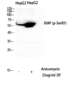
- Western Blot analysis of 293 cells using XIAP Polyclonal Antibody diluted at 1:1000. Secondary antibody(catalog#:RS0002) was diluted at 1:20000
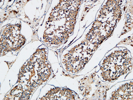
- Immunohistochemical analysis of paraffin-embedded Human testis. 1, Antibody was diluted at 1:200(4° overnight). 2, High-pressure and temperature EDTA, pH8.0 was used for antigen retrieval. 3,Secondary antibody was diluted at 1:200(room temperature, 30min).
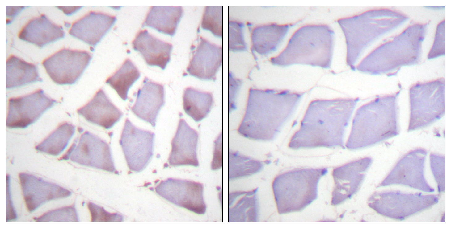
- Immunohistochemistry analysis of paraffin-embedded human skeletal muscle tissue, using XIAP Antibody. The picture on the right is blocked with the synthesized peptide.
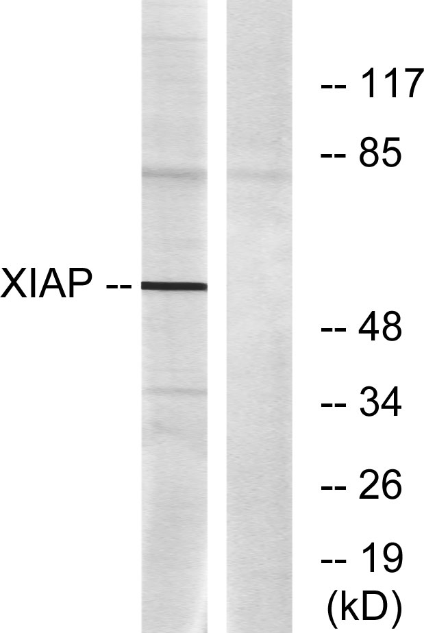
- Western blot analysis of lysates from 293 cells, using XIAP Antibody. The lane on the right is blocked with the synthesized peptide.