SIRP-α1 Polyclonal Antibody
- Catalog No.:YT4301
- Applications:WB;IHC;IF;ELISA
- Reactivity:Human;Mouse;Rat
- Target:
- SHPS1
- Fields:
- >>Osteoclast differentiation
- Gene Name:
- SIRPA
- Protein Name:
- Tyrosine-protein phosphatase non-receptor type substrate 1
- Human Gene Id:
- 140885
- Human Swiss Prot No:
- P78324
- Mouse Gene Id:
- 19261
- Mouse Swiss Prot No:
- P97797
- Rat Gene Id:
- 25528
- Rat Swiss Prot No:
- P97710
- Immunogen:
- The antiserum was produced against synthesized peptide derived from human Sirp alpha1. AA range:451-500
- Specificity:
- SIRP-α1 Polyclonal Antibody detects endogenous levels of SIRP-α1 protein.
- Formulation:
- Liquid in PBS containing 50% glycerol, 0.5% BSA and 0.02% sodium azide.
- Source:
- Polyclonal, Rabbit,IgG
- Dilution:
- WB 1:500 - 1:2000. IHC 1:100 - 1:300. ELISA: 1:20000.. IF 1:50-200
- Purification:
- The antibody was affinity-purified from rabbit antiserum by affinity-chromatography using epitope-specific immunogen.
- Concentration:
- 1 mg/ml
- Storage Stability:
- -15°C to -25°C/1 year(Do not lower than -25°C)
- Other Name:
- SIRPA;BIT;MFR;MYD1;PTPNS1;SHPS1;SIRP;Tyrosine-protein phosphatase non-receptor type substrate 1;SHP substrate 1;SHPS-1;Brain Ig-like molecule with tyrosine-based activation motifs;Bit;CD172 antigen-like family member A;Inhibito
- Observed Band(KD):
- 55kD
- Background:
- The protein encoded by this gene is a member of the signal-regulatory-protein (SIRP) family, and also belongs to the immunoglobulin superfamily. SIRP family members are receptor-type transmembrane glycoproteins known to be involved in the negative regulation of receptor tyrosine kinase-coupled signaling processes. This protein can be phosphorylated by tyrosine kinases. The phospho-tyrosine residues of this PTP have been shown to recruit SH2 domain containing tyrosine phosphatases (PTP), and serve as substrates of PTPs. This protein was found to participate in signal transduction mediated by various growth factor receptors. CD47 has been demonstrated to be a ligand for this receptor protein. This gene and its product share very high similarity with several other members of the SIRP family. These related genes are located in close proximity to each other on chromosome 20p13. Multiple alternati
- Function:
- function:Immunoglobulin-like cell surface receptor for CD47. Acts as docking protein and induces translocation of PTPN6, PTPN11 and other binding partners from the cytosol to the plasma membrane. Supports adhesion of cerebellar neurons, neurite outgrowth and glial cell attachment. May play a key role in intracellular signaling during synaptogenesis and in synaptic function (By similarity). Involved in the negative regulation of receptor tyrosine kinase-coupled cellular responses induced by cell adhesion, growth factors or insulin. Mediates negative regulation of phagocytosis, mast cell activation and dendritic cell activation. CD47 binding prevents maturation of immature dendritic cells and inhibits cytokine production by mature dendritic cells.,PTM:N-glycosylated.,PTM:Phosphorylated on tyrosine residues in response to stimulation with EGF, growth hormone, insulin and PDGF. Dephosphoryla
- Subcellular Location:
- Membrane; Single-pass type I membrane protein.
- Expression:
- Ubiquitous. Highly expressed in brain. Detected on myeloid cells, but not T-cells. Detected at lower levels in heart, placenta, lung, testis, ovary, colon, liver, small intestine, prostate, spleen, kidney, skeletal muscle and pancreas.
Suppression of the hyaluronic acid pathway induces M1 macrophages polarization via STAT1 in glioblastoma. Cell Death Discovery2022 Apr;8(1):1-13. Human U937 monocytes,U251 cell,LN229 cell
- June 19-2018
- WESTERN IMMUNOBLOTTING PROTOCOL
- June 19-2018
- IMMUNOHISTOCHEMISTRY-PARAFFIN PROTOCOL
- June 19-2018
- IMMUNOFLUORESCENCE PROTOCOL
- September 08-2020
- FLOW-CYTOMEYRT-PROTOCOL
- May 20-2022
- Cell-Based ELISA│解您多样本WB检测之困扰
- July 13-2018
- CELL-BASED-ELISA-PROTOCOL-FOR-ACETYL-PROTEIN
- July 13-2018
- CELL-BASED-ELISA-PROTOCOL-FOR-PHOSPHO-PROTEIN
- July 13-2018
- Antibody-FAQs
- Products Images

- Western Blot analysis of various cells using SIRP-α1 Polyclonal Antibody
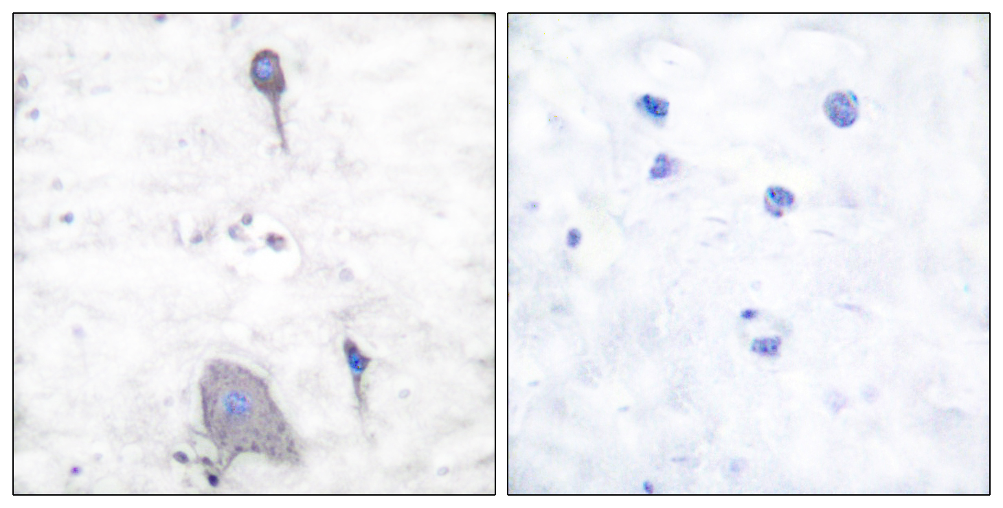
- Immunohistochemistry analysis of paraffin-embedded human brain tissue, using Sirp alpha1 Antibody. The picture on the right is blocked with the synthesized peptide.
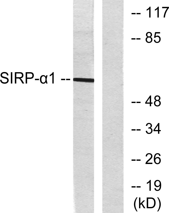
- Western blot analysis of lysates from HepG2 cells, using Sirp alpha1 Antibody. The lane on the right is blocked with the synthesized peptide.


