Rad23B Polyclonal Antibody
- Catalog No.:YT3962
- Applications:WB;IHC;IF;ELISA
- Reactivity:Human;Mouse;Rat
- Target:
- Rad23B
- Fields:
- >>Nucleotide excision repair;>>Protein processing in endoplasmic reticulum
- Gene Name:
- RAD23B
- Protein Name:
- UV excision repair protein RAD23 homolog B
- Human Gene Id:
- 5887
- Human Swiss Prot No:
- P54727
- Mouse Gene Id:
- 19359
- Mouse Swiss Prot No:
- P54728
- Rat Gene Id:
- 298012
- Rat Swiss Prot No:
- Q4KMA2
- Immunogen:
- The antiserum was produced against synthesized peptide derived from human RAD23B. AA range:1-50
- Specificity:
- Rad23B Polyclonal Antibody detects endogenous levels of Rad23B protein.
- Formulation:
- Liquid in PBS containing 50% glycerol, 0.5% BSA and 0.02% sodium azide.
- Source:
- Polyclonal, Rabbit,IgG
- Dilution:
- WB 1:500 - 1:2000. IHC 1:100 - 1:300. IF 1:200 - 1:1000. ELISA: 1:20000. Not yet tested in other applications.
- Purification:
- The antibody was affinity-purified from rabbit antiserum by affinity-chromatography using epitope-specific immunogen.
- Concentration:
- 1 mg/ml
- Storage Stability:
- -15°C to -25°C/1 year(Do not lower than -25°C)
- Other Name:
- RAD23B;UV excision repair protein RAD23 homolog B;HR23B;hHR23B;XP-C repair-complementing complex 58 kDa protein;p58
- Observed Band(KD):
- 58kD
- Background:
- The protein encoded by this gene is one of two human homologs of Saccharomyces cerevisiae Rad23, a protein involved in the nucleotide excision repair (NER). This protein was found to be a component of the protein complex that specifically complements the NER defect of xeroderma pigmentosum group C (XP-c) cell extracts in vitro. This protein was also shown to interact with, and elevate the nucleotide excision activity of 3-methyladenine-DNA glycosylase (MPG), which suggested a role in DNA damage recognition in base excision repair. This protein contains an N-terminal ubiquitin-like domain, which was reported to interact with 26S proteasome, and thus this protein may be involved in the ubiquitin mediated proteolytic pathway in cells. Alternative splicing results in multiple transcript variants encoding distinct isoforms. [provided by RefSeq, Sep 2011],
- Function:
- domain:The ubiquitin-like domain mediates interaction with MJD.,function:Plays a central role both in proteosomal degradation of misfolded proteins and DNA repair. Central component of a complex required to couple deglycosylation and proteasome-mediated degradation of misfolded proteins in the endoplasmic reticulun that are retrotranslocated in the cytosol. Involved in DNA excision repair by stabilizing XPC protein. May play a part in DNA damage recognition and/or in altering chromatin structure to allow access by damage-processing enzymes.,similarity:Belongs to the RAD23 family.,similarity:Contains 1 STI1 domain.,similarity:Contains 1 ubiquitin-like domain.,similarity:Contains 2 UBA domains.,subunit:Component of a complex required to couple retrotranslocation, ubiquitination and deglycosylation composed of NGLY1, SAKS1, AMFR, VCP and RAD23B (By similarity). Interacts with the 26S protea
- Subcellular Location:
- Nucleus. Cytoplasm. The intracellular distribution is cell cycle dependent. Localized to the nucleus and the cytoplasm during G1 phase. Nuclear levels decrease during S-phase; upon entering mitosis, relocalizes in the cytoplasm without association with chromatin.
- Expression:
- Epithelium,Testis,Thymus,Uterus,
- June 19-2018
- WESTERN IMMUNOBLOTTING PROTOCOL
- June 19-2018
- IMMUNOHISTOCHEMISTRY-PARAFFIN PROTOCOL
- June 19-2018
- IMMUNOFLUORESCENCE PROTOCOL
- September 08-2020
- FLOW-CYTOMEYRT-PROTOCOL
- May 20-2022
- Cell-Based ELISA│解您多样本WB检测之困扰
- July 13-2018
- CELL-BASED-ELISA-PROTOCOL-FOR-ACETYL-PROTEIN
- July 13-2018
- CELL-BASED-ELISA-PROTOCOL-FOR-PHOSPHO-PROTEIN
- July 13-2018
- Antibody-FAQs
- Products Images
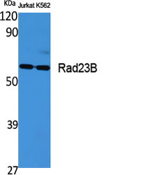
- Western Blot analysis of various cells using Rad23B Polyclonal Antibody
.jpg)
- Western Blot analysis of HepG2 cells using Rad23B Polyclonal Antibody
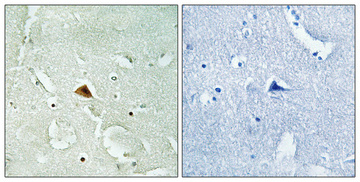
- Immunohistochemical analysis of paraffin-embedded Human brain. Antibody was diluted at 1:100(4° overnight). High-pressure and temperature Tris-EDTA,pH8.0 was used for antigen retrieval. Negetive contrl (right) obtaned from antibody was pre-absorbed by immunogen peptide.
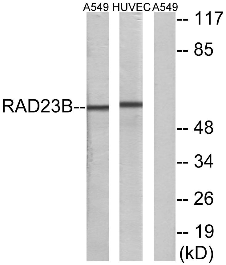
- Western blot analysis of lysates from A549 and HUVEC cells, using RAD23B Antibody. The lane on the right is blocked with the synthesized peptide.


