S100A9 (Phospho Thr113) rabbit pAb
- Catalog No.:YP1699
- Applications:WB
- Reactivity:Human;Mouse;Rat
- Target:
- S100A9
- Fields:
- >>IL-17 signaling pathway
- Gene Name:
- S100A9 CAGB CFAG MRP14
- Protein Name:
- S100A9 (Phospho-Thr113)
- Human Gene Id:
- 6280
- Human Swiss Prot No:
- P06702
- Mouse Gene Id:
- 20202
- Mouse Swiss Prot No:
- P31725
- Rat Gene Id:
- 94195
- Rat Swiss Prot No:
- P50116
- Immunogen:
- Synthesized peptide derived from human S100A9 (Phospho-Thr113)
- Specificity:
- This antibody detects endogenous levels of S100A9 (Phospho-Thr113) at Human, Mouse,Rat
- Formulation:
- Liquid in PBS containing 50% glycerol, 0.5% BSA and 0.02% sodium azide.
- Source:
- Polyclonal, Rabbit,IgG
- Dilution:
- WB 1:500-2000
- Purification:
- The antibody was affinity-purified from rabbit serum by affinity-chromatography using specific immunogen.
- Concentration:
- 1 mg/ml
- Storage Stability:
- -15°C to -25°C/1 year(Do not lower than -25°C)
- Other Name:
- Protein S100-A9 (Calgranulin-B) (Calprotectin L1H subunit) (Leukocyte L1 complex heavy chain) (Migration inhibitory factor-related protein 14) (MRP-14) (p14) (S100 calcium-binding protein A9)
- Molecular Weight(Da):
- 13kD
- Background:
- S100 calcium binding protein A9(S100A9) Homo sapiens The protein encoded by this gene is a member of the S100 family of proteins containing 2 EF-hand calcium-binding motifs. S100 proteins are localized in the cytoplasm and/or nucleus of a wide range of cells, and involved in the regulation of a number of cellular processes such as cell cycle progression and differentiation. S100 genes include at least 13 members which are located as a cluster on chromosome 1q21. This protein may function in the inhibition of casein kinase and altered expression of this protein is associated with the disease cystic fibrosis. This antimicrobial protein exhibits antifungal and antibacterial activity. [provided by RefSeq, Nov 2014],
- Function:
- function:Expressed by macrophages in acutely inflammated tissues and in chronic inflammations. Seem to be an inhibitor of protein kinases. Also expressed in epithelial cells constitutively or induced during dermatoses. May interact with components of the intermediate filaments in monocytes and epithelial cells.,miscellaneous:Has been shown to bind calcium.,similarity:Belongs to the S-100 family.,similarity:Contains 2 EF-hand domains.,subunit:Interacts with CEACAM3 in a calcium-dependent manner.,
- Subcellular Location:
- Secreted. Cytoplasm . Cytoplasm, cytoskeleton . Cell membrane ; Peripheral membrane protein . Predominantly localized in the cytoplasm. Upon elevation of the intracellular calcium level, translocated from the cytoplasm to the cytoskeleton and the cell membrane (PubMed:18786929). Upon neutrophil activation or endothelial adhesion of monocytes, is secreted via a microtubule-mediated, alternative pathway (PubMed:15598812). .
- Expression:
- Calprotectin (S100A8/9) is predominantly expressed in myeloid cells. Except for inflammatory conditions, the expression is restricted to a specific stage of myeloid differentiation since both proteins are expressed in circulating neutrophils and monocytes but are absent in normal tissue macrophages and lymphocytes. Under chronic inflammatory conditions, such as psoriasis and malignant disorders, also expressed in the epidermis. Found in high concentrations at local sites of inflammation or in the serum of patients with inflammatory diseases such as rheumatoid, cystic fibrosis, inflammatory bowel disease, Crohn's disease, giant cell arteritis, cystic fibrosis, Sjogren's syndrome, systemic lupus erythematosus, and progressive systemic sclerosis. Involved in the formation and deposition of am
- June 19-2018
- WESTERN IMMUNOBLOTTING PROTOCOL
- June 19-2018
- IMMUNOHISTOCHEMISTRY-PARAFFIN PROTOCOL
- June 19-2018
- IMMUNOFLUORESCENCE PROTOCOL
- September 08-2020
- FLOW-CYTOMEYRT-PROTOCOL
- May 20-2022
- Cell-Based ELISA│解您多样本WB检测之困扰
- July 13-2018
- CELL-BASED-ELISA-PROTOCOL-FOR-ACETYL-PROTEIN
- July 13-2018
- CELL-BASED-ELISA-PROTOCOL-FOR-PHOSPHO-PROTEIN
- July 13-2018
- Antibody-FAQs
- Products Images

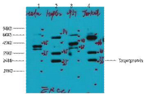
- Western Blot analysis of 1 A549 cell, 2 LPS 100ng/mL 30min treated ,using primary antibody at 1:1000 dilution. Secondary antibody(catalog#:RS23920) was diluted at 1:10000


