FKHR (Phospho Ser249) rabbit pAb
- Catalog No.:YP1671
- Applications:WB
- Reactivity:Human;Mouse;Rat
- Target:
- FoxO1
- Fields:
- >>FoxO signaling pathway;>>AMPK signaling pathway;>>Longevity regulating pathway;>>Longevity regulating pathway - multiple species;>>Cellular senescence;>>Insulin signaling pathway;>>Thyroid hormone signaling pathway;>>Glucagon signaling pathway;>>Insulin resistance;>>AGE-RAGE signaling pathway in diabetic complications;>>Alcoholic liver disease;>>Shigellosis;>>Human papillomavirus infection;>>Pathways in cancer;>>Transcriptional misregulation in cancer;>>Prostate cancer
- Gene Name:
- FOXO1 FKHR FOXO1A
- Protein Name:
- FKHR (Phospho-Ser249)
- Human Gene Id:
- 2308
- Human Swiss Prot No:
- Q12778
- Mouse Gene Id:
- 56458
- Mouse Swiss Prot No:
- Q9R1E0
- Rat Gene Id:
- 84482
- Rat Swiss Prot No:
- G3V7R4
- Immunogen:
- Synthesized peptide derived from human FKHR (Phospho-Ser249)
- Specificity:
- This antibody detects endogenous levels of FKHR (Phospho-Ser249) at Human, Mouse,Rat
- Formulation:
- Liquid in PBS containing 50% glycerol, 0.5% BSA and 0.02% sodium azide.
- Source:
- Polyclonal, Rabbit,IgG
- Dilution:
- WB 1:500-2000
- Purification:
- The antibody was affinity-purified from rabbit serum by affinity-chromatography using specific immunogen.
- Concentration:
- 1 mg/ml
- Storage Stability:
- -15°C to -25°C/1 year(Do not lower than -25°C)
- Other Name:
- Forkhead box protein O1 (Forkhead box protein O1A) (Forkhead in rhabdomyosarcoma)
- Molecular Weight(Da):
- 72kD
- Background:
- This gene belongs to the forkhead family of transcription factors which are characterized by a distinct forkhead domain. The specific function of this gene has not yet been determined; however, it may play a role in myogenic growth and differentiation. Translocation of this gene with PAX3 has been associated with alveolar rhabdomyosarcoma. [provided by RefSeq, Jul 2008],
- Function:
- disease:Chromosomal aberrations involving FOXO1 are a cause of rhabdomyosarcoma 2 (RMS2) [MIM:268220]; also known as alveolar rhabdomyosarcoma. Translocation (2;13)(q35;q14) with PAX3; translocation t(1;13)(p36;q14) with PAX7. The resulting protein is a transcriptional activator.,function:Transcription factor.,PTM:Phosphorylated by AKT1; insulin-induced (By similarity). IGF1 rapidly induces phosphorylation of Ser-256, Thr-24, and Ser-319. Phosphorylation of Ser-256 decreases DNA-binding activity and promotes the phosphorylation of Thr-24, and Ser-319, permitting phosphorylation of Ser-322 and Ser-325, probably by CK1, leading to nuclear exclusion and loss of function. Phosphorylation of Ser-329 is independent of IGF1 and leads to reduced function. Phosphorylated upon DNA damage, probably by ATM or ATR.,similarity:Contains 1 fork-head DNA-binding domain.,subcellular location:Shuttles betw
- Subcellular Location:
- Cytoplasm . Nucleus . Shuttles between the cytoplasm and nucleus. Largely nuclear in unstimulated cells (PubMed:11311120, PubMed:12228231, PubMed:19221179, PubMed:21245099, PubMed:20543840, PubMed:25009184). In osteoblasts, colocalizes with ATF4 and RUNX2 in the nucleus (By similarity). Serum deprivation increases localization to the nucleus, leading to activate expression of SOX9 and subsequent chondrogenesis (By similarity). Insulin-induced phosphorylation at Ser-256 by PKB/AKT1 leads, via stimulation of Thr-24 phosphorylation, to binding of 14-3-3 proteins and nuclear export to the cytoplasm where it is degraded by the ubiquitin-proteosomal pathway (PubMed:11237865, PubMed:12228231). Phosphorylation at Ser-249 by CDK1 disrupts binding of 14-3-3 proteins and promotes nuclear accumulation
- Expression:
- Ubiquitous.
- June 19-2018
- WESTERN IMMUNOBLOTTING PROTOCOL
- June 19-2018
- IMMUNOHISTOCHEMISTRY-PARAFFIN PROTOCOL
- June 19-2018
- IMMUNOFLUORESCENCE PROTOCOL
- September 08-2020
- FLOW-CYTOMEYRT-PROTOCOL
- May 20-2022
- Cell-Based ELISA│解您多样本WB检测之困扰
- July 13-2018
- CELL-BASED-ELISA-PROTOCOL-FOR-ACETYL-PROTEIN
- July 13-2018
- CELL-BASED-ELISA-PROTOCOL-FOR-PHOSPHO-PROTEIN
- July 13-2018
- Antibody-FAQs
- Products Images

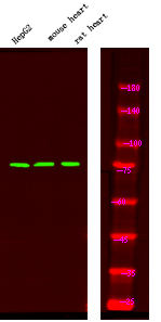
- Western Blot analysis of HepG2 mouse brain tissue, rat brain tissue, using primary antibody at 1:1000 dilution 4°C, overnight. Secondary antibody(catalog#:RS23920) was diluted at 1:10000 25°C,1.5hours


