NFAT1 (Phospho Ser326) rabbit pAb
- Catalog No.:YP1563
- Applications:WB;ELISA;IHC
- Reactivity:Human;Mouse;Rat
- Target:
- NFAT1
- Fields:
- >>cGMP-PKG signaling pathway;>>Cellular senescence;>>Wnt signaling pathway;>>Axon guidance;>>VEGF signaling pathway;>>Osteoclast differentiation;>>C-type lectin receptor signaling pathway;>>Natural killer cell mediated cytotoxicity;>>Th1 and Th2 cell differentiation;>>Th17 cell differentiation;>>T cell receptor signaling pathway;>>B cell receptor signaling pathway;>>Oxytocin signaling pathway;>>Yersinia infection;>>Hepatitis B;>>Human cytomegalovirus infection;>>Human T-cell leukemia virus 1 infection;>>Kaposi sarcoma-associated herpesvirus infection;>>Human immunodeficiency virus 1 infection;>>PD-L1 expression and PD-1 checkpoint pathway in cancer;>>Lipid and atherosclerosis
- Gene Name:
- NFATC2 NFAT1 NFATP
- Protein Name:
- NFAT1 (Phospho Ser326)
- Human Gene Id:
- 4773
- Human Swiss Prot No:
- Q13469
- Mouse Gene Id:
- 18019
- Mouse Swiss Prot No:
- Q60591
- Immunogen:
- Synthesized peptide derived from human NFAT1 (Phospho Ser326)
- Specificity:
- This antibody detects endogenous levels of Human,Mouse,Rat NFAT1 (Phospho Ser326)
- Formulation:
- Liquid in PBS containing 50% glycerol, 0.5% BSA and 0.02% sodium azide.
- Source:
- Polyclonal, Rabbit,IgG
- Dilution:
- WB 1:500-2000;IHC 1:50-300; ELISA 2000-20000
- Purification:
- The antibody was affinity-purified from rabbit serum by affinity-chromatography using specific immunogen.
- Concentration:
- 1 mg/ml
- Storage Stability:
- -15°C to -25°C/1 year(Do not lower than -25°C)
- Other Name:
- Nuclear factor of activated T-cells, cytoplasmic 2 (NF-ATc2;NFATc2;NFAT pre-existing subunit;NF-ATp;T-cell transcription factor NFAT1)
- Observed Band(KD):
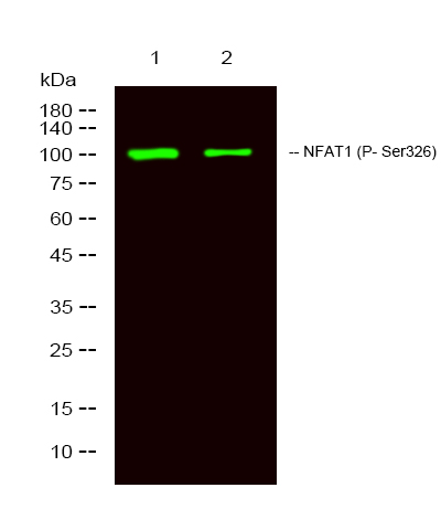
- 100kD
- Background:
- This gene is a member of the nuclear factor of activated T cells (NFAT) family. The product of this gene is a DNA-binding protein with a REL-homology region (RHR) and an NFAT-homology region (NHR). This protein is present in the cytosol and only translocates to the nucleus upon T cell receptor (TCR) stimulation, where it becomes a member of the nuclear factors of activated T cells transcription complex. This complex plays a central role in inducing gene transcription during the immune response. Alternate transcriptional splice variants encoding different isoforms have been characterized. [provided by RefSeq, Apr 2012],
- Function:
- alternative products:Additional isoforms seem to exist,domain:Rel Similarity Domain (RSD) allows DNA-binding and cooperative interactions with AP1 factors.,function:Plays a role in the inducible expression of cytokine genes in T-cells, especially in the induction of the IL-2, IL-3, IL-4, TNF-alpha or GM-CSF.,induction:Inducibly expressed in T-lymphocytes upon activation of the T-cell receptor (TCR) complex. Induced after co-addition of phorbol 12-myristate 13-acetate (PMA) and ionomycin.,PTM:In resting cells, phosphorylated by NFATC-kinase on at least 18 sites in the 99-363 region. Upon cell stimulation, all these sites except Ser-243 are dephosphorylated by calcineurin. Dephosphorylation induces a conformational change that simultaneously exposes an NLS and masks an NES, which results in nuclear localization. Simultaneously, Ser-53 or Ser-56 is phosphorylated; which is required for full
- Subcellular Location:
- Cytoplasm. Nucleus. Cytoplasmic for the phosphorylated form and nuclear after activation that is controlled by calcineurin-mediated dephosphorylation. Rapid nuclear exit of NFATC is thought to be one mechanism by which cells distinguish between sustained and transient calcium signals. The subcellular localization of NFATC plays a key role in the regulation of gene transcription.
- Expression:
- Expressed in thymus, spleen, heart, testis, brain, placenta, muscle and pancreas. Isoform 1 is highly expressed in the small intestine, heart, testis, prostate, thymus, placenta and thyroid. Isoform 3 is highly expressed in stomach, uterus, placenta, trachea and thyroid.
TRPA1 deficiency attenuates cardiac fibrosis via regulating GRK5/NFAT signaling in diabetic rats. Yao Xu WB Rat cardiac tissue
- June 19-2018
- WESTERN IMMUNOBLOTTING PROTOCOL
- June 19-2018
- IMMUNOHISTOCHEMISTRY-PARAFFIN PROTOCOL
- June 19-2018
- IMMUNOFLUORESCENCE PROTOCOL
- September 08-2020
- FLOW-CYTOMEYRT-PROTOCOL
- May 20-2022
- Cell-Based ELISA│解您多样本WB检测之困扰
- July 13-2018
- CELL-BASED-ELISA-PROTOCOL-FOR-ACETYL-PROTEIN
- July 13-2018
- CELL-BASED-ELISA-PROTOCOL-FOR-PHOSPHO-PROTEIN
- July 13-2018
- Antibody-FAQs
- Products Images

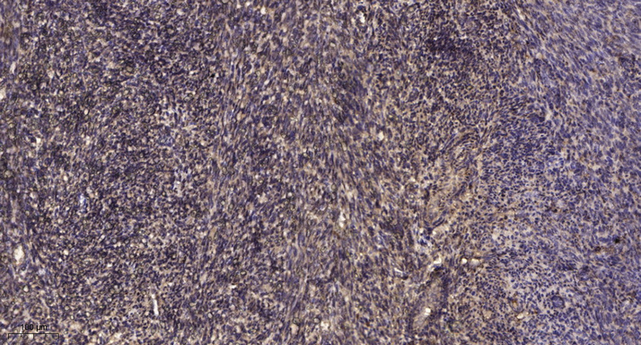
- Immunohistochemical analysis of paraffin-embedded human uterus. 1, Antibody was diluted at 1:200(4° overnight). 2, Tris-EDTA,pH9.0 was used for antigen retrieval. 3,Secondary antibody was diluted at 1:200(room temperature, 45min).

- Western Blot analysis of 1 MCF-7 treated with LPS, 2 MCF-7,using primary antibody at 1:1000 dilution. Secondary antibody(catalog#:RS23920) was diluted at 1:10000


