Peroxiredoxin 1 Monoclonal Antibody(8E7)
- Catalog No.:YM3112
- Applications:WB;IHC;IF
- Reactivity:Human;Mouse;Rat
- Target:
- Prdx1
- Fields:
- >>Peroxisome;>>Amoebiasis
- Gene Name:
- PRDX1
- Protein Name:
- Peroxiredoxin-1
- Human Gene Id:
- 5052
- Human Swiss Prot No:
- Q06830
- Mouse Gene Id:
- 18477
- Mouse Swiss Prot No:
- P35700
- Rat Gene Id:
- 117254
- Rat Swiss Prot No:
- Q63716
- Immunogen:
- Recombinant Protein of Peroxiredoxin-1
- Specificity:
- The antibody detects endogenous Peroxiredoxin 1 protein.
- Formulation:
- PBS, pH 7.4, containing 0.5%BSA, 0.02% sodium azide as Preservative and 50% Glycerol.
- Source:
- Monoclonal, Mouse
- Dilution:
- WB 1:1000-3000 IF 1:100-200 IHC 1:50-300
- Purification:
- The antibody was affinity-purified from mouse ascites by affinity-chromatography using specific immunogen.
- Storage Stability:
- -15°C to -25°C/1 year(Do not lower than -25°C)
- Other Name:
- PRDX1;PAGA;PAGB;TDPX2;Peroxiredoxin-1;Natural killer cell-enhancing factor A;NKEF-A;Proliferation-associated gene protein;PAG;Thioredoxin peroxidase 2;Thioredoxin-dependent peroxide reductase 2
- Observed Band(KD):
- 21kD
- Background:
- This gene encodes a member of the peroxiredoxin family of antioxidant enzymes, which reduce hydrogen peroxide and alkyl hydroperoxides. The encoded protein may play an antioxidant protective role in cells, and may contribute to the antiviral activity of CD8(+) T-cells. This protein may have a proliferative effect and play a role in cancer development or progression. Four transcript variants encoding the same protein have been identified for this gene. [provided by RefSeq, Jan 2011],
- Function:
- catalytic activity:2 R'-SH + ROOH = R'-S-S-R' + H(2)O + ROH.,function:Involved in redox regulation of the cell. Reduces peroxides with reducing equivalents provided through the thioredoxin system but not from glutaredoxin. May play an important role in eliminating peroxides generated during metabolism. Might participate in the signaling cascades of growth factors and tumor necrosis factor-alpha by regulating the intracellular concentrations of H(2)O(2).,induction:Constitutively expressed in most human cells; is induced to higher levels upon serum stimulation in untransformed and transformed cells.,miscellaneous:Inactivated upon oxidative stress by overoxidation of Cys-52 to Cys-SO(2)H and Cys-SO(3)H. Cys-SO(2)H is retroreduced to Cys-SOH after removal of H(2)O(2), while Cys-SO(3)H may be irreversibly oxidized.,miscellaneous:The active site is the redox-active Cys-52 oxidized to Cys-SOH.
- Subcellular Location:
- Cytoplasm . Melanosome . Identified by mass spectrometry in melanosome fractions from stage I to stage IV.
- Expression:
- Brain,Cajal-Retzius cell,Fetal brain cortex,Urinary bladder,
- June 19-2018
- WESTERN IMMUNOBLOTTING PROTOCOL
- June 19-2018
- IMMUNOHISTOCHEMISTRY-PARAFFIN PROTOCOL
- June 19-2018
- IMMUNOFLUORESCENCE PROTOCOL
- September 08-2020
- FLOW-CYTOMEYRT-PROTOCOL
- May 20-2022
- Cell-Based ELISA│解您多样本WB检测之困扰
- July 13-2018
- CELL-BASED-ELISA-PROTOCOL-FOR-ACETYL-PROTEIN
- July 13-2018
- CELL-BASED-ELISA-PROTOCOL-FOR-PHOSPHO-PROTEIN
- July 13-2018
- Antibody-FAQs
- Products Images
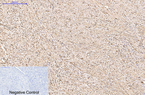
- Immunohistochemical analysis of paraffin-embedded Human-uterus-cancer tissue. 1,Peroxiredoxin 1 Monoclonal Antibody(8E7) was diluted at 1:200(4°C,overnight). 2, Sodium citrate pH 6.0 was used for antibody retrieval(>98°C,20min). 3,Secondary antibody was diluted at 1:200(room tempeRature, 30min). Negative control was used by secondary antibody only.
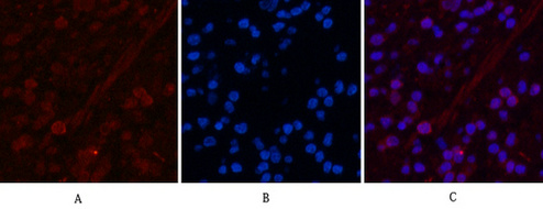
- Immunofluorescence analysis of Human-appendix tissue. 1,Peroxiredoxin 1 Monoclonal Antibody(8E7)(red) was diluted at 1:200(4°C,overnight). 2, Cy3 labled Secondary antibody was diluted at 1:300(room temperature, 50min).3, Picture B: DAPI(blue) 10min. Picture A:Target. Picture B: DAPI. Picture C: merge of A+B
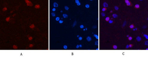
- Immunofluorescence analysis of Rat-brain tissue. 1,Peroxiredoxin 1 Monoclonal Antibody(8E7)(red) was diluted at 1:200(4°C,overnight). 2, Cy3 labled Secondary antibody was diluted at 1:300(room temperature, 50min).3, Picture B: DAPI(blue) 10min. Picture A:Target. Picture B: DAPI. Picture C: merge of A+B
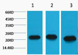
- Western blot analysis of 1) MCF7, 2) Rat Kidney Tissue, 3) Mouse Brain Tissue, diluted at 1:2000.
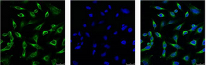
- IF analysis of Hela with antibody (Left) and DAPI (Right) diluted at 1:100.


