PRC1(Phospho Thr481)Polyclonal Antibody
- Catalog No.:YP1213
- Applications:WB;IHC
- Reactivity:Human;Rat;Mouse;
- Target:
- PRC1(Phospho-Thr481)Polyclonal Antibody
- Gene Name:
- PRC1
- Protein Name:
- Protein regulator of cytokinesis 1
- Human Gene Id:
- 9055
- Human Swiss Prot No:
- O43663
- Mouse Swiss Prot No:
- Q99K43
- Immunogen:
- Synthetic Phospho peptide from human protein at AA range: 481
- Specificity:
- The antibody detects endogenous PRC1Phospho occurs at Thr481
- Formulation:
- Liquid in PBS containing 50% glycerol, 0.5% BSA and 0.02% sodium azide.
- Source:
- Polyclonal, Rabbit,IgG
- Dilution:
- WB 1:500-2000;IHC 1:50-300
- Purification:
- The antibody was affinity-purified from rabbit antiserum by affinity-chromatography using epitope-specific immunogen.
- Concentration:
- 1 mg/ml
- Storage Stability:
- -15°C to -25°C/1 year(Do not lower than -25°C)
- Other Name:
- Protein regulator of cytokinesis 1
- Observed Band(KD):
- 72kD
- Background:
- This gene encodes a protein that is involved in cytokinesis. The protein is present at high levels during the S and G2/M phases of mitosis but its levels drop dramatically when the cell exits mitosis and enters the G1 phase. It is located in the nucleus during interphase, becomes associated with mitotic spindles in a highly dynamic manner during mitosis, and localizes to the cell mid-body during cytokinesis. This protein has been shown to be a substrate of several cyclin-dependent kinases (CDKs). It is necessary for polarizing parallel microtubules and concentrating the factors responsible for contractile ring assembly. Alternative splicing results in multiple transcript variants. [provided by RefSeq, Jun 2012],
- Function:
- function:KIF4A translocates PRC1 to the plus ends of interdigitating spindle microtubules during the metaphase to anaphase transition, an essential step for the formation of an organized central spindle midzone and midbody and for successful cytokinesis. Required for KIF14 localization to the central spindle and midbody. Acts as a microtubule-binding and bundling protein both in vivo and vitro. May function as an in vivo cyclin-CDK substrate.,PTM:Phosphorylated; very weak in G1/S phase cells. Much higher levels of phosphorylation are detected at later cell cycle phases, reaching a maximum during mitosis.,similarity:Belongs to the MAP65/ASE1 family.,subcellular location:Predominantly localized to the nucleus of interphase cells. During mitosis becomes associated with the mitotic spindle poles and localizes with the cell midbody during cytokinesis.,subunit:Interacts with the C-terminal Rho
- Subcellular Location:
- Nucleus . Cytoplasm. Cytoplasm, cytoskeleton, spindle pole . Midbody . Chromosome . Colocalized with KIF20B in the nucleus of bladder carcinoma cells at the interphase. Colocalized with KIF20B in bladder carcinoma cells at prophase, metaphase, early anaphase, at the midzone in late anaphase and at the contractile ring in telophase (PubMed:17409436). Predominantly localized to the nucleus of interphase cells. During mitosis becomes associated with the mitotic spindle poles and localizes with the cell midbody during cytokinesis. Co-localizes with PRC1 in early mitosis and at the spindle midzone from anaphase B to telophase (PubMed:15297875, PubMed:15625105). .
- Expression:
- Overexpressed in bladder cancer cells (PubMed:17409436).
- June 19-2018
- WESTERN IMMUNOBLOTTING PROTOCOL
- June 19-2018
- IMMUNOHISTOCHEMISTRY-PARAFFIN PROTOCOL
- June 19-2018
- IMMUNOFLUORESCENCE PROTOCOL
- September 08-2020
- FLOW-CYTOMEYRT-PROTOCOL
- May 20-2022
- Cell-Based ELISA│解您多样本WB检测之困扰
- July 13-2018
- CELL-BASED-ELISA-PROTOCOL-FOR-ACETYL-PROTEIN
- July 13-2018
- CELL-BASED-ELISA-PROTOCOL-FOR-PHOSPHO-PROTEIN
- July 13-2018
- Antibody-FAQs
- Products Images
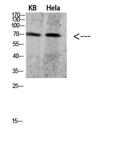
- Western blot analysis of 293T VEC lysate, antibody was diluted at 500. Secondary antibody(catalog#:RS0002) was diluted at 1:20000
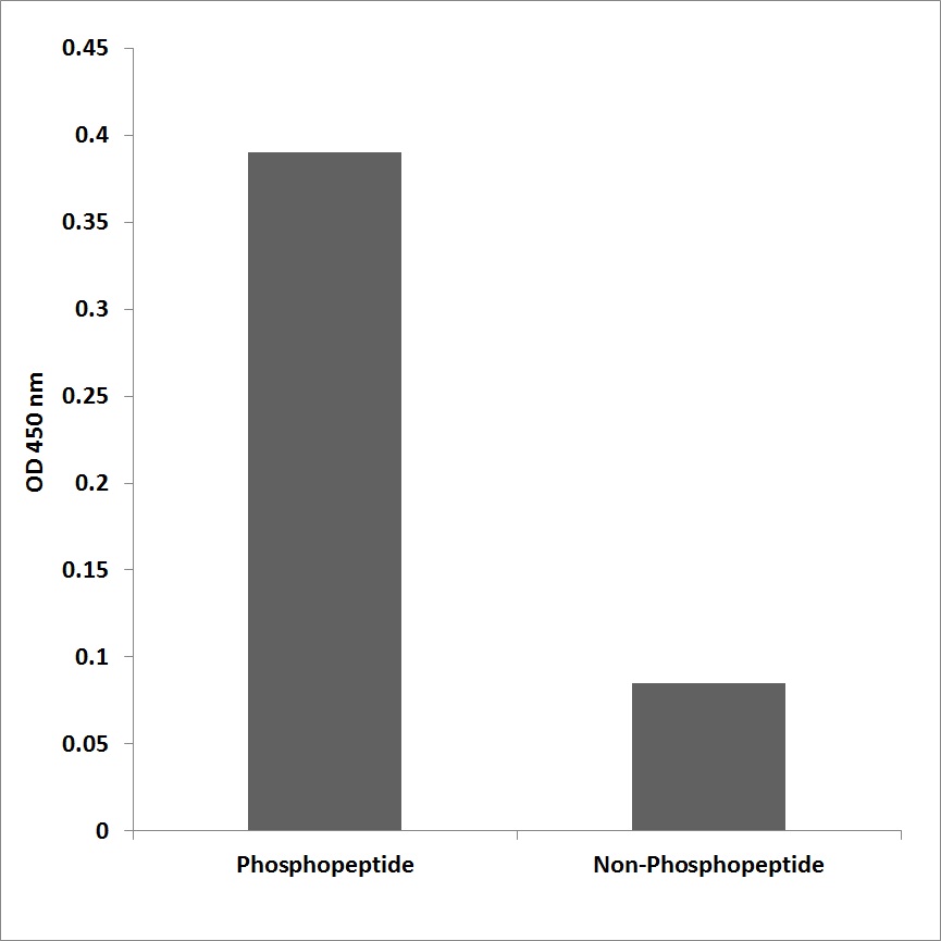
- Enzyme-Linked Immunosorbent Assay (Phospho-ELISA) for Immunogen Phosphopeptide (Phospho-left) and Non-Phosphopeptide (Phospho-right), using PRC1 (Phospho-Thr481) Antibody
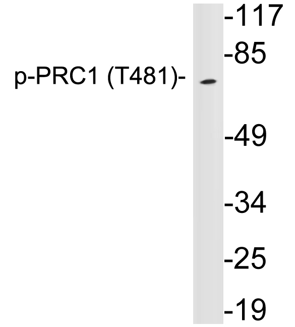
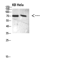
- Western blot analysis of lysates from HeLa cells, using p-PRC1 (Phospho-Thr481) antibody.
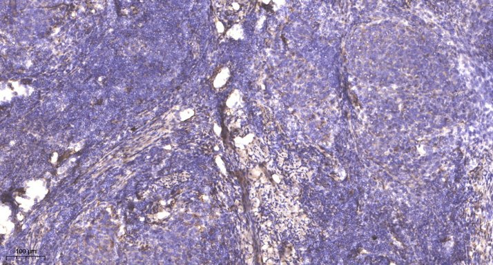
- Immunohistochemical analysis of paraffin-embedded human cervical carcinoma. 1, Antibody was diluted at 1:200(4° overnight). 2, Tris-EDTA,pH9.0 was used for antigen retrieval. 3,Secondary antibody was diluted at 1:200(room temperature, 45min).