GSDME N-terminal rabbit pAb
- Catalog No.:YT7990
- Applications:IF;WB;IHC
- Reactivity:Human;Mouse;Rat
- Target:
- GSDME
- Gene Name:
- GSDME
- Protein Name:
- Gasdermin-E,DFNA5,Dfna5h,EG14210,2310037D07Rik .4932441K13Rik ,ICERE 1,ICERE-1,Deafness, autosomal dominant 5,Non-syndromic hearing impairment protein 5,GSDME-NT
- Human Swiss Prot No:
- O60443
- Immunogen:
- Synthesized peptide derived from human GSDME. AA range:1-100
- Specificity:
- The antibody detects endogenous full lenth and n-ternal fragment of gsdme protein,
- Formulation:
- Liquid in PBS containing 50% glycerol, and 0.02% sodium azide.
- Source:
- Polyclonal, Rabbit,IgG
- Dilution:
- IF 1:50-200 WB 1:5000-20000 IHC 1:200-300
- Purification:
- The antibody was affinity-purified from rabbit antiserum by affinity-chromatography using epitope-specific immunogen.
- Concentration:
- 1 mg/ml
- Storage Stability:
- -15°C to -25°C/1 year(Do not lower than -25°C)
- Observed Band(KD):
- 36 55kD
- Function:
- Cleavage at Asp-270 by CASP3 (mature and uncleaved precursor forms) or granzyme B (GZMB) relieves autoinhibition and is sufficient to initiate pyroptosis.
- Subcellular Location:
- [Gasdermin-E, N-terminal]: Cell membrane ; Multi-pass membrane protein .; [Gasdermin-E]: Cytoplasm, cytosol .
- Expression:
- Expressed in cochlea (PubMed:9771715). Low level of expression in heart, brain, placenta, lung, liver, skeletal muscle, kidney and pancreas, with highest expression in placenta (PubMed:9771715).
- June 19-2018
- WESTERN IMMUNOBLOTTING PROTOCOL
- June 19-2018
- IMMUNOHISTOCHEMISTRY-PARAFFIN PROTOCOL
- June 19-2018
- IMMUNOFLUORESCENCE PROTOCOL
- September 08-2020
- FLOW-CYTOMEYRT-PROTOCOL
- May 20-2022
- Cell-Based ELISA│解您多样本WB检测之困扰
- July 13-2018
- CELL-BASED-ELISA-PROTOCOL-FOR-ACETYL-PROTEIN
- July 13-2018
- CELL-BASED-ELISA-PROTOCOL-FOR-PHOSPHO-PROTEIN
- July 13-2018
- Antibody-FAQs
- Products Images
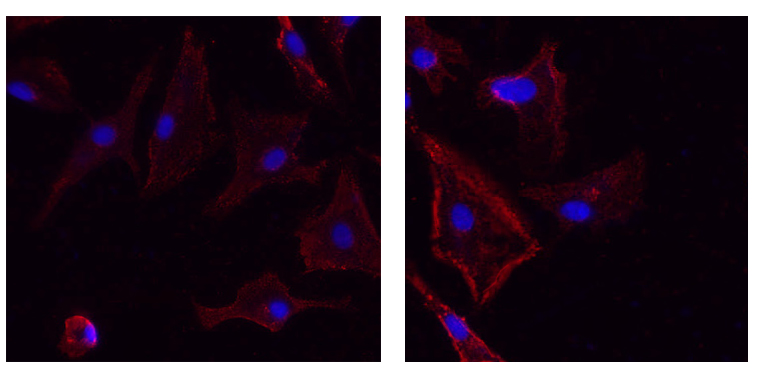
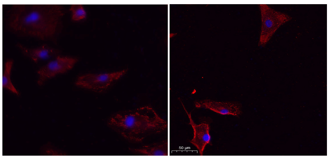
- Immunofluorescence analysis of un-treated (left) A549 and UV treated(right) A549 cell. 1,primary Antibody was diluted at 1:200(4°C overnight). 2, Goat Anti Rabbit IgG (H&L) - Alexa Fluor 594 Secondary antibody was diluted at 1:1000(room temperature, 50min).3, Picture B: DAPI(blue) 10min.
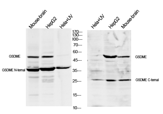
- Western blot analysis of lysates from 1)mouse-brain, 2)Hela cells treated by UV 15min,3) HepG2 cells, primary antibody was diluted at 1:1000, 4°over night, secondary antibody HRP goat anti rabbit (Immunoway:RS0002) was diluted at 1:10000