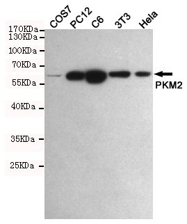
PKM2 Polyclonal Antibody
- Catalog No.:YT3777
- Applications:IF;WB;IHC;ELISA
- Reactivity:Human;Mouse;Rat
- Target:
- PKM2
- Fields:
- >>Glycolysis / Gluconeogenesis;>>Pyruvate metabolism;>>Metabolic pathways;>>Carbon metabolism;>>Biosynthesis of amino acids;>>Glucagon signaling pathway;>>Type II diabetes mellitus;>>Human papillomavirus infection;>>Viral carcinogenesis;>>Central carbon metabolism in cancer
- Gene Name:
- PKM
- Protein Name:
- Pyruvate kinase isozymes M1/M2
- Human Gene Id:
- 5315
- Human Swiss Prot No:
- P14618
- Mouse Gene Id:
- 18746
- Mouse Swiss Prot No:
- P52480
- Rat Gene Id:
- 25630
- Rat Swiss Prot No:
- P11980
- Immunogen:
- The antiserum was produced against synthesized peptide derived from human PKM2. AA range:181-230
- Specificity:
- PKM2 Polyclonal Antibody detects endogenous levels of PKM2 protein.
- Formulation:
- Liquid in PBS containing 50% glycerol, 0.5% BSA and 0.02% sodium azide.
- Source:
- Polyclonal, Rabbit,IgG
- Dilution:
- IF 1:50-200 WB 1:500-2000, ELISA 1:10000-20000 IHC 1:50-300
- Purification:
- The antibody was affinity-purified from rabbit antiserum by affinity-chromatography using epitope-specific immunogen.
- Concentration:
- 1 mg/ml
- Storage Stability:
- -15°C to -25°C/1 year(Do not lower than -25°C)
- Other Name:
- PKM;OIP3;PK2;PK3;PKM2;Pyruvate kinase isozymes M1/M2;Cytosolic thyroid hormone-binding protein;CTHBP;Opa-interacting protein 3;OIP-3;Pyruvate kinase 2/3;Pyruvate kinase muscle isozyme;Thyroid hormone-binding protein 1;THBP1;Tu
- Observed Band(KD):
- 58kD
- Background:
- This gene encodes a protein involved in glycolysis. The encoded protein is a pyruvate kinase that catalyzes the transfer of a phosphoryl group from phosphoenolpyruvate to ADP, generating ATP and pyruvate. This protein has been shown to interact with thyroid hormone and may mediate cellular metabolic effects induced by thyroid hormones. This protein has been found to bind Opa protein, a bacterial outer membrane protein involved in gonococcal adherence to and invasion of human cells, suggesting a role of this protein in bacterial pathogenesis. Several alternatively spliced transcript variants encoding a few distinct isoforms have been reported. [provided by RefSeq, May 2011],
- Function:
- catalytic activity:ATP + pyruvate = ADP + phosphoenolpyruvate.,cofactor:Divalent metal cations.,cofactor:Magnesium.,cofactor:Potassium.,enzyme regulation:Isoform M2 is allosterically activated by D-fructose 1,6-biphosphate (FBP). Inhibited by oxalate and 3,3',5-triiodo-L-thyronine (T3).,function:Glycolytic enzyme that catalyzes the transfer of a phosphoryl group from phosphoenolpyruvate (PEP) to ADP, generating ATP.,miscellaneous:There are 4 isozymes of pyruvate kinase in mammals: L, R, M1 and M2. L type is major isozyme in the liver, R is found in red cells, M1 is the main form in muscle, heart and brain, and M2 is found in early fetal tissues as well as in most cancer cells.,online information:Pyruvate kinase entry,pathway:Carbohydrate degradation; glycolysis; pyruvate from D-glyceraldehyde 3-phosphate: step 5/5.,PTM:Phosphorylated upon DNA damage, probably by ATM or ATR.,similarity:Be
- Subcellular Location:
- [Isoform M2]: Cytoplasm . Nucleus . Translocates to the nucleus in response to various signals, such as EGF receptor activation or apoptotic stimuli (PubMed:17308100, PubMed:22056988, PubMed:24120661). Nuclear translocation is promoted by acetylation by EP300 (PubMed:24120661). Deacetylation by SIRT6 promotes its nuclear export in a process dependent of XPO4, thereby suppressing its ability to activate transcription and promote tumorigenesis (PubMed:26787900). .; [Isoform M1]: Cytoplasm .
- Expression:
- [Isoform M2]: Specifically expressed in proliferating cells, such as embryonic stem cells, embryonic carcinoma cells, as well as cancer cells. ; [Isoform M1]: Expressed in adult tissues (PubMed:18337823). Not expressed in tumor cells (PubMed:18337823).
- June 19-2018
- WESTERN IMMUNOBLOTTING PROTOCOL
- June 19-2018
- IMMUNOHISTOCHEMISTRY-PARAFFIN PROTOCOL
- June 19-2018
- IMMUNOFLUORESCENCE PROTOCOL
- September 08-2020
- FLOW-CYTOMEYRT-PROTOCOL
- May 20-2022
- Cell-Based ELISA│解您多样本WB检测之困扰
- July 13-2018
- CELL-BASED-ELISA-PROTOCOL-FOR-ACETYL-PROTEIN
- July 13-2018
- CELL-BASED-ELISA-PROTOCOL-FOR-PHOSPHO-PROTEIN
- July 13-2018
- Antibody-FAQs