Synapsin I Polyclonal Antibody
- Catalog No.:YT4483
- Applications:WB;IHC;IF;ELISA
- Reactivity:Human;Mouse;Rat
- Target:
- Synapsin I
- Gene Name:
- SYN1
- Protein Name:
- Synapsin-1
- Human Gene Id:
- 6853
- Human Swiss Prot No:
- P17600
- Mouse Gene Id:
- 20964
- Mouse Swiss Prot No:
- O88935
- Rat Gene Id:
- 24949
- Rat Swiss Prot No:
- P09951
- Immunogen:
- The antiserum was produced against synthesized peptide derived from human Synapsin. AA range:3-52
- Specificity:
- Synapsin I Polyclonal Antibody detects endogenous levels of Synapsin I protein.
- Formulation:
- Liquid in PBS containing 50% glycerol, 0.5% BSA and 0.02% sodium azide.
- Source:
- Polyclonal, Rabbit,IgG
- Dilution:
- WB 1:500 - 1:2000. IHC 1:100 - 1:300. IF 1:200 - 1:1000. ELISA: 1:20000. Not yet tested in other applications.
- Purification:
- The antibody was affinity-purified from rabbit antiserum by affinity-chromatography using epitope-specific immunogen.
- Concentration:
- 1 mg/ml
- Storage Stability:
- -15°C to -25°C/1 year(Do not lower than -25°C)
- Other Name:
- SYN1;Synapsin-1;Brain protein 4.1;Synapsin I
- Observed Band(KD):
- 74kD
- Background:
- This gene is a member of the synapsin gene family. Synapsins encode neuronal phosphoproteins which associate with the cytoplasmic surface of synaptic vesicles. Family members are characterized by common protein domains, and they are implicated in synaptogenesis and the modulation of neurotransmitter release, suggesting a potential role in several neuropsychiatric diseases. This member of the synapsin family plays a role in regulation of axonogenesis and synaptogenesis. The protein encoded serves as a substrate for several different protein kinases and phosphorylation may function in the regulation of this protein in the nerve terminal. Mutations in this gene may be associated with X-linked disorders with primary neuronal degeneration such as Rett syndrome. Alternatively spliced transcript variants encoding different isoforms have been identified. [provided by RefSeq, Jul 2008],
- Function:
- disease:Defects in SYN1 are a cause of epilepsy X-linked with variable learning disabilities and behavior disorders [MIM:300491]. XELBD is characterized by variable combinations of epilepsy, learning difficulties, macrocephaly, and aggressive behavior.,function:Neuronal phosphoprotein that coats synaptic vesicles, binds to the cytoskeleton, and is believed to function in the regulation of neurotransmitter release. The complex formed with NOS1 and CAPON proteins is necessary for specific nitric-oxid functions at a presynaptic level.,PTM:Substrate of at least four different protein kinases. It is probable that phosphorylation plays a role in the regulation of synapsin-1 in the nerve terminal. Phosphorylated upon DNA damage, probably by ATM or ATR.,similarity:Belongs to the synapsin family.,subunit:Homodimer. Interacts with CAPON. Forms a ternary complex with NOS1. Isoform Ib interacts with
- Subcellular Location:
- Cell junction, synapse. Golgi apparatus .
- Expression:
- Brain,Brain cortex,
- June 19-2018
- WESTERN IMMUNOBLOTTING PROTOCOL
- June 19-2018
- IMMUNOHISTOCHEMISTRY-PARAFFIN PROTOCOL
- June 19-2018
- IMMUNOFLUORESCENCE PROTOCOL
- September 08-2020
- FLOW-CYTOMEYRT-PROTOCOL
- May 20-2022
- Cell-Based ELISA│解您多样本WB检测之困扰
- July 13-2018
- CELL-BASED-ELISA-PROTOCOL-FOR-ACETYL-PROTEIN
- July 13-2018
- CELL-BASED-ELISA-PROTOCOL-FOR-PHOSPHO-PROTEIN
- July 13-2018
- Antibody-FAQs
- Products Images
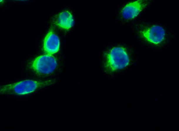
- Immunofluorescence analysis of Hela cell. 1,Synapsin I Polyclonal Antibody(green) was diluted at 1:200(4° overnight). 2, Goat Anti Rabbit Alexa Fluor 488 Catalog:RS3211 was diluted at 1:1000(room temperature, 50min). 3 DAPI(blue) 10min.
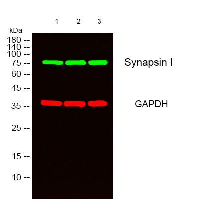
- Western blot analysis of lysates from 1) Hela, 2) 293, 3) NIH-3T3 cells, (Green) primary antibody was diluted at 1:1000, 4°over night, secondary antibody(cat:RS23920)was diluted at 1:10000, 37° 1hour. (Red) GAPDH Monoclonal Antibody(2B8) (cat:YM3029) antibody was diluted at 1:5000 as loading control, 4° over night,secondary antibody(cat:RS23710)was diluted at 1:10000, 37° 1hour.
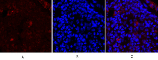
- Immunofluorescence analysis of rat-lung tissue. 1,Synapsin I Polyclonal Antibody(red) was diluted at 1:200(4°C,overnight). 2, Cy3 labled Secondary antibody was diluted at 1:300(room temperature, 50min).3, Picture B: DAPI(blue) 10min. Picture A:Target. Picture B: DAPI. Picture C: merge of A+B
- Immunofluorescence analysis of rat-lung tissue. 1,Synapsin I Polyclonal Antibody(red) was diluted at 1:200(4°C,overnight). 2, Cy3 labled Secondary antibody was diluted at 1:300(room temperature, 50min).3, Picture B: DAPI(blue) 10min. Picture A:Target. Picture B: DAPI. Picture C: merge of A+B
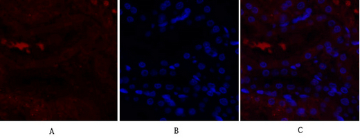
- Immunofluorescence analysis of rat-kidney tissue. 1,Synapsin I Polyclonal Antibody(red) was diluted at 1:200(4°C,overnight). 2, Cy3 labled Secondary antibody was diluted at 1:300(room temperature, 50min).3, Picture B: DAPI(blue) 10min. Picture A:Target. Picture B: DAPI. Picture C: merge of A+B
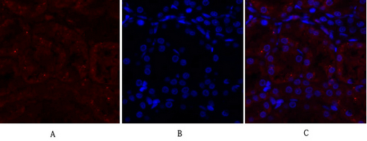
- Immunofluorescence analysis of rat-kidney tissue. 1,Synapsin I Polyclonal Antibody(red) was diluted at 1:200(4°C,overnight). 2, Cy3 labled Secondary antibody was diluted at 1:300(room temperature, 50min).3, Picture B: DAPI(blue) 10min. Picture A:Target. Picture B: DAPI. Picture C: merge of A+B

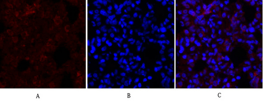
- Immunofluorescence analysis of rat-spleen tissue. 1,Synapsin I Polyclonal Antibody(red) was diluted at 1:200(4°C,overnight). 2, Cy3 labled Secondary antibody was diluted at 1:300(room temperature, 50min).3, Picture B: DAPI(blue) 10min. Picture A:Target. Picture B: DAPI. Picture C: merge of A+B

- Immunofluorescence analysis of rat-spleen tissue. 1,Synapsin I Polyclonal Antibody(red) was diluted at 1:200(4°C,overnight). 2, Cy3 labled Secondary antibody was diluted at 1:300(room temperature, 50min).3, Picture B: DAPI(blue) 10min. Picture A:Target. Picture B: DAPI. Picture C: merge of A+B
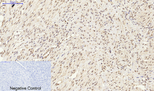
- Immunohistochemical analysis of paraffin-embedded Human-uterus-cancer tissue. 1,Synapsin I Polyclonal Antibody was diluted at 1:200(4°C,overnight). 2, Sodium citrate pH 6.0 was used for antibody retrieval(>98°C,20min). 3,Secondary antibody was diluted at 1:200(room tempeRature, 30min). Negative control was used by secondary antibody only.
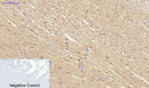
- Immunohistochemical analysis of paraffin-embedded Rat-heart tissue. 1,Synapsin I Polyclonal Antibody was diluted at 1:200(4°C,overnight). 2, Sodium citrate pH 6.0 was used for antibody retrieval(>98°C,20min). 3,Secondary antibody was diluted at 1:200(room tempeRature, 30min). Negative control was used by secondary antibody only.
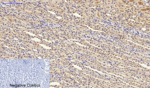
- Immunohistochemical analysis of paraffin-embedded Rat-kidney tissue. 1,Synapsin I Polyclonal Antibody was diluted at 1:200(4°C,overnight). 2, Sodium citrate pH 6.0 was used for antibody retrieval(>98°C,20min). 3,Secondary antibody was diluted at 1:200(room tempeRature, 30min). Negative control was used by secondary antibody only.
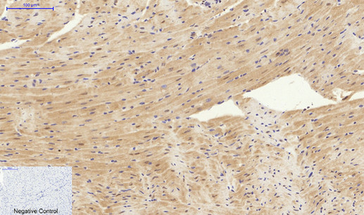
- Immunohistochemical analysis of paraffin-embedded Mouse-heart tissue. 1,Synapsin I Polyclonal Antibody was diluted at 1:200(4°C,overnight). 2, Sodium citrate pH 6.0 was used for antibody retrieval(>98°C,20min). 3,Secondary antibody was diluted at 1:200(room tempeRature, 30min). Negative control was used by secondary antibody only.