Raf-1 Polyclonal Antibody
- Catalog No.:YT3979
- Applications:WB;IHC;IF;ELISA
- Reactivity:Human;Mouse;Rat
- Target:
- Raf-1
- Fields:
- >>EGFR tyrosine kinase inhibitor resistance;>>Endocrine resistance;>>MAPK signaling pathway;>>ErbB signaling pathway;>>Ras signaling pathway;>>Rap1 signaling pathway;>>cGMP-PKG signaling pathway;>>cAMP signaling pathway;>>Chemokine signaling pathway;>>FoxO signaling pathway;>>Sphingolipid signaling pathway;>>Phospholipase D signaling pathway;>>Autophagy - animal;>>mTOR signaling pathway;>>PI3K-Akt signaling pathway;>>Apoptosis;>>Cellular senescence;>>Vascular smooth muscle contraction;>>Axon guidance;>>VEGF signaling pathway;>>Apelin signaling pathway;>>Focal adhesion;>>Gap junction;>>Signaling pathways regulating pluripotency of stem cells;>>Neutrophil extracellular trap formation;>>C-type lectin receptor signaling pathway;>>JAK-STAT signaling pathway;>>Natural killer cell mediated cytotoxicity;>>T cell receptor signaling pathway;>>B cell receptor signaling pathway;>>Fc epsilon RI signaling pathway;>>Fc gamma R-mediated phagocytosis;>>Long-term potentiation;>>Neurotrophin signaling pa
- Gene Name:
- RAF1
- Protein Name:
- RAF proto-oncogene serine/threonine-protein kinase
- Human Gene Id:
- 5894
- Human Swiss Prot No:
- P04049
- Mouse Gene Id:
- 110157
- Mouse Swiss Prot No:
- Q99N57
- Rat Gene Id:
- 24703
- Rat Swiss Prot No:
- P11345
- Immunogen:
- The antiserum was produced against synthesized peptide derived from human C-RAF. AA range:11-60
- Specificity:
- Raf-1 Polyclonal Antibody detects endogenous levels of Raf-1 protein.
- Formulation:
- Liquid in PBS containing 50% glycerol, 0.5% BSA and 0.02% sodium azide.
- Source:
- Polyclonal, Rabbit,IgG
- Dilution:
- WB 1:500 - 1:2000. IHC 1:100 - 1:300. ELISA: 1:5000.. IF 1:50-200
- Purification:
- The antibody was affinity-purified from rabbit antiserum by affinity-chromatography using epitope-specific immunogen.
- Concentration:
- 1 mg/ml
- Storage Stability:
- -15°C to -25°C/1 year(Do not lower than -25°C)
- Other Name:
- RAF1;RAF;RAF proto-oncogene serine/threonine-protein kinase;Proto-oncogene c-RAF;cRaf;Raf-1
- Observed Band(KD):
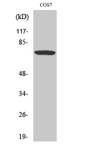
- 73kD
- Background:
- This gene is the cellular homolog of viral raf gene (v-raf). The encoded protein is a MAP kinase kinase kinase (MAP3K), which functions downstream of the Ras family of membrane associated GTPases to which it binds directly. Once activated, the cellular RAF1 protein can phosphorylate to activate the dual specificity protein kinases MEK1 and MEK2, which in turn phosphorylate to activate the serine/threonine specific protein kinases, ERK1 and ERK2. Activated ERKs are pleiotropic effectors of cell physiology and play an important role in the control of gene expression involved in the cell division cycle, apoptosis, cell differentiation and cell migration. Mutations in this gene are associated with Noonan syndrome 5 and LEOPARD syndrome 2. [provided by RefSeq, Jul 2008],
- Function:
- catalytic activity:ATP + a protein = ADP + a phosphoprotein.,cofactor:Binds 2 zinc ions per subunit.,disease:Defects in RAF1 are the cause of LEOPARD syndrome type 2 (LEOPARD syndrome-2) [MIM:611554]. LEOPARD syndrome is an autosomal dominant disorder allelic with Noonan syndrome. The acronym LEOPARD stands for lentigines, electrocardiographic conduction abnormalities, ocular hypertelorism, pulmonic stenosis, abnormalities of genitalia, retardation of growth, and deafness.,disease:Defects in RAF1 are the cause of Noonan syndrome type 5 (NS5) [MIM:611553]. Noonan syndrome (NS) is a disorder characterized by dysmorphic facial features, short stature, hypertelorism, cardiac anomalies, deafness, motor delay, and a bleeding diathesis. It is a genetically heterogeneous and relatively common syndrome, with an estimated incidence of 1 in 1000-2500 live births.,function:Involved in the transducti
- Subcellular Location:
- Cytoplasm. Cell membrane. Mitochondrion. Nucleus. Colocalizes with RGS14 and BRAF in both the cytoplasm and membranes. Phosphorylation at Ser-259 impairs its membrane accumulation. Recruited to the cell membrane by the active Ras protein. Phosphorylation at Ser-338 and Ser-339 by PAK1 is required for its mitochondrial localization. Retinoic acid-induced Ser-621 phosphorylated form of RAF1 is predominantly localized at the nucleus.
- Expression:
- In skeletal muscle, isoform 1 is more abundant than isoform 2.
The oncoprotein HBXIP enhances migration of breast cancer cells through increasing filopodia formation involving MEKK2/ERK1/2/Capn4 signaling. CANCER LETTERS 2014 Oct 07 WB Human MCF-7 cell
Four new antitumor metabolites isolated from a mutant 3-f-31 strain derived from Penicillium purpurogenum G59. EUROPEAN JOURNAL OF MEDICINAL CHEMISTRY 2018 Sep 08 WB Human HCT-116 cell
Effects of hydroxysafflor yellow A on rats with collagen-induced arthritis. BIOCHEMICAL AND BIOPHYSICAL RESEARCH COMMUNICATIONS Biochem Bioph Res Co. 2021 Sep;570:26 WB Rat 1:500 Synovial
Nanocarrier of Pin1 inhibitor based on supercritical fluid technology inhibits cancer metastasis by blocking multiple signaling pathways Regenerative Biomaterials Dayun Yang WB Human HuH7 cell,HepG2 cell
Ambient NO2 hinders neutrophil extracellular trap formation in rats: Assessment of the role of neutrophil autophagy. Fang Xiao WB Rat 1:1000 neutrophils
- June 19-2018
- WESTERN IMMUNOBLOTTING PROTOCOL
- June 19-2018
- IMMUNOHISTOCHEMISTRY-PARAFFIN PROTOCOL
- June 19-2018
- IMMUNOFLUORESCENCE PROTOCOL
- September 08-2020
- FLOW-CYTOMEYRT-PROTOCOL
- May 20-2022
- Cell-Based ELISA│解您多样本WB检测之困扰
- July 13-2018
- CELL-BASED-ELISA-PROTOCOL-FOR-ACETYL-PROTEIN
- July 13-2018
- CELL-BASED-ELISA-PROTOCOL-FOR-PHOSPHO-PROTEIN
- July 13-2018
- Antibody-FAQs
- Products Images

- Ambient NO2 hinders neutrophil extracellular trap formation in rats: Assessment of the role of neutrophil autophagy. Fang Xiao WB Rat 1:1000 neutrophils

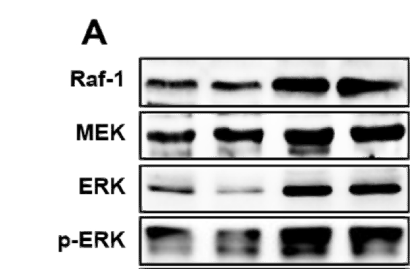
- Western blot analysis in MCF-7 cells transfected with siRaf-1. Cancer Letters 355 (2014) 288–296

- Western Blot analysis of various cells using Raf-1 Polyclonal Antibody diluted at 1:2000

- The picture was kindly provided by our customer

- Immunohistochemistry analysis of paraffin-embedded human brain tissue, using C-RAF Antibody. The picture on the right is blocked with the synthesized peptide.

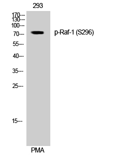
- Western blot analysis of lysates from 293 cells, treated with PMA 125ng/ml 30', using C-RAF Antibody. The lane on the right is blocked with the synthesized peptide.


