PIP5KIII Polyclonal Antibody
- Catalog No.:YT3734
- Applications:WB;IHC
- Reactivity:Human;Mouse
- Target:
- PIP5K
- Fields:
- >>Inositol phosphate metabolism;>>Metabolic pathways;>>Phosphatidylinositol signaling system;>>Phagosome;>>Regulation of actin cytoskeleton
- Gene Name:
- PIKFYVE
- Protein Name:
- 1-phosphatidylinositol 3-phosphate 5-kinase
- Human Gene Id:
- 200576
- Human Swiss Prot No:
- Q9Y2I7
- Mouse Gene Id:
- 18711
- Mouse Swiss Prot No:
- Q9Z1T6
- Immunogen:
- The antiserum was produced against synthesized peptide derived from human PIP5K. AA range:71-120
- Specificity:
- PIP5KIII Polyclonal Antibody detects endogenous levels of PIP5KIII protein.
- Formulation:
- Liquid in PBS containing 50% glycerol, 0.5% BSA and 0.02% sodium azide.
- Source:
- Polyclonal, Rabbit,IgG
- Dilution:
- WB 1:500-2000;IHC 1:50-300
- Purification:
- The antibody was affinity-purified from rabbit antiserum by affinity-chromatography using epitope-specific immunogen.
- Concentration:
- 1 mg/ml
- Storage Stability:
- -15°C to -25°C/1 year(Do not lower than -25°C)
- Other Name:
- PIKFYVE;KIAA0981;PIP5K3;1-phosphatidylinositol 3-phosphate 5-kinase;Phosphatidylinositol 3-phosphate 5-kinase;FYVE finger-containing phosphoinositide kinase;PIKfyve;Phosphatidylinositol 3-phosphate 5-kinase type III;PIPkin-III;Type
- Observed Band(KD):
- 237kD
- Background:
- Phosphorylated derivatives of phosphatidylinositol (PtdIns) regulate cytoskeletal functions, membrane trafficking, and receptor signaling by recruiting protein complexes to cell- and endosomal-membranes. Humans have multiple PtdIns proteins that differ by the degree and position of phosphorylation of the inositol ring. This gene encodes an enzyme (PIKfyve; also known as phosphatidylinositol-3-phosphate 5-kinase type III or PIPKIII) that phosphorylates the D-5 position in PtdIns and phosphatidylinositol-3-phosphate (PtdIns3P) to make PtdIns5P and PtdIns(3,5)biphosphate. The D-5 position also can be phosphorylated by type I PtdIns4P-5-kinases (PIP5Ks) that are encoded by distinct genes and preferentially phosphorylate D-4 phosphorylated PtdIns. In contrast, PIKfyve preferentially phosphorylates D-3 phosphorylated PtdIns. In addition to being a lipid kinase, PIKf
- Function:
- catalytic activity:ATP + 1-phosphatidyl-1D-myo-inositol 4-phosphate = ADP + 1-phosphatidyl-1D-myo-inositol 4,5-bisphosphate.,disease:Defects in PIKFYVE are the cause of corneal fleck dystrophy (CFD) [MIM:121850]. CFD is an autosomal dominant disorder of the cornea characterized by numerous small white flecks scattered in all levels of the stroma. Although CFD may occasionally cause mild photophobia, patients are typically asymptomatic and have normal vision.,function:Supports the intracellular PIP pool and to a lesser extent, the PI 4,5-P(2) pool. It generates PIP from PI and, to a lesser extent, PI 4,5-P(2) from PI 4-P. There are indications that it phosphorylates the D-5 rather than the D-4 position. Has a role in endosome-related membrane trafficking.,similarity:Contains 1 DEP domain.,similarity:Contains 1 FYVE-type zinc finger.,similarity:Contains 1 PI5K domain.,subcellular location:
- Subcellular Location:
- Endosome membrane ; Peripheral membrane protein . Early endosome membrane ; Peripheral membrane protein. Cytoplasmic vesicle, phagosome membrane ; Peripheral membrane protein . Late endosome membrane ; Peripheral membrane protein . Mainly associated with membranes of the late endocytic pathway. .
- Expression:
- Brain,Epithelium,PCR rescued clones,T-cell,
- June 19-2018
- WESTERN IMMUNOBLOTTING PROTOCOL
- June 19-2018
- IMMUNOHISTOCHEMISTRY-PARAFFIN PROTOCOL
- June 19-2018
- IMMUNOFLUORESCENCE PROTOCOL
- September 08-2020
- FLOW-CYTOMEYRT-PROTOCOL
- May 20-2022
- Cell-Based ELISA│解您多样本WB检测之困扰
- July 13-2018
- CELL-BASED-ELISA-PROTOCOL-FOR-ACETYL-PROTEIN
- July 13-2018
- CELL-BASED-ELISA-PROTOCOL-FOR-PHOSPHO-PROTEIN
- July 13-2018
- Antibody-FAQs
- Products Images
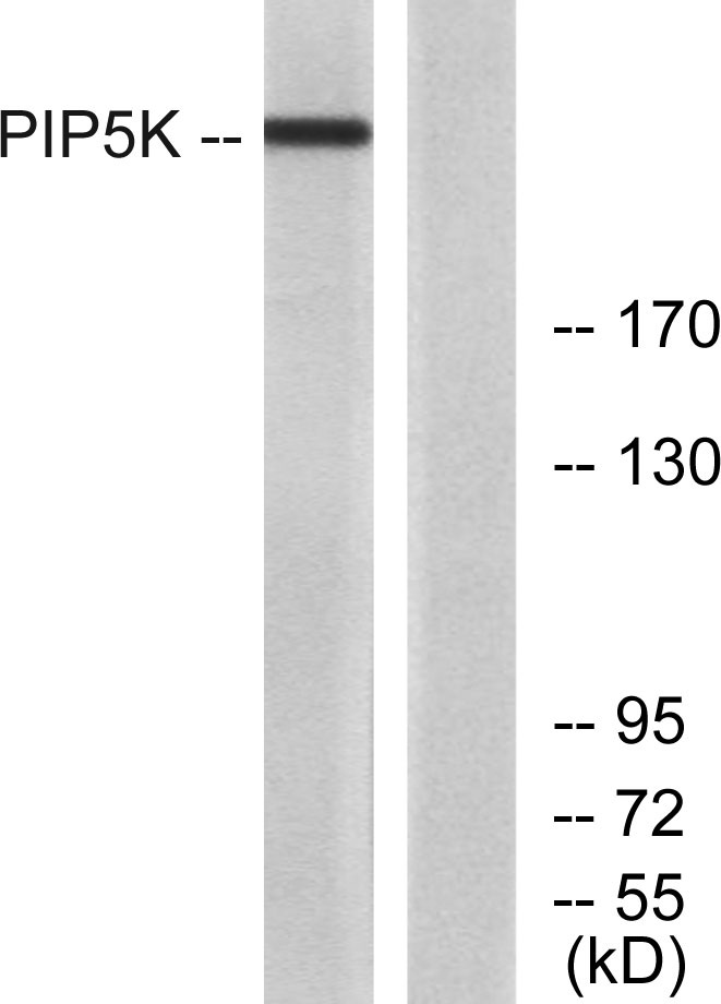
- Western Blot analysis of various cells using PIP5KIII Polyclonal Antibody
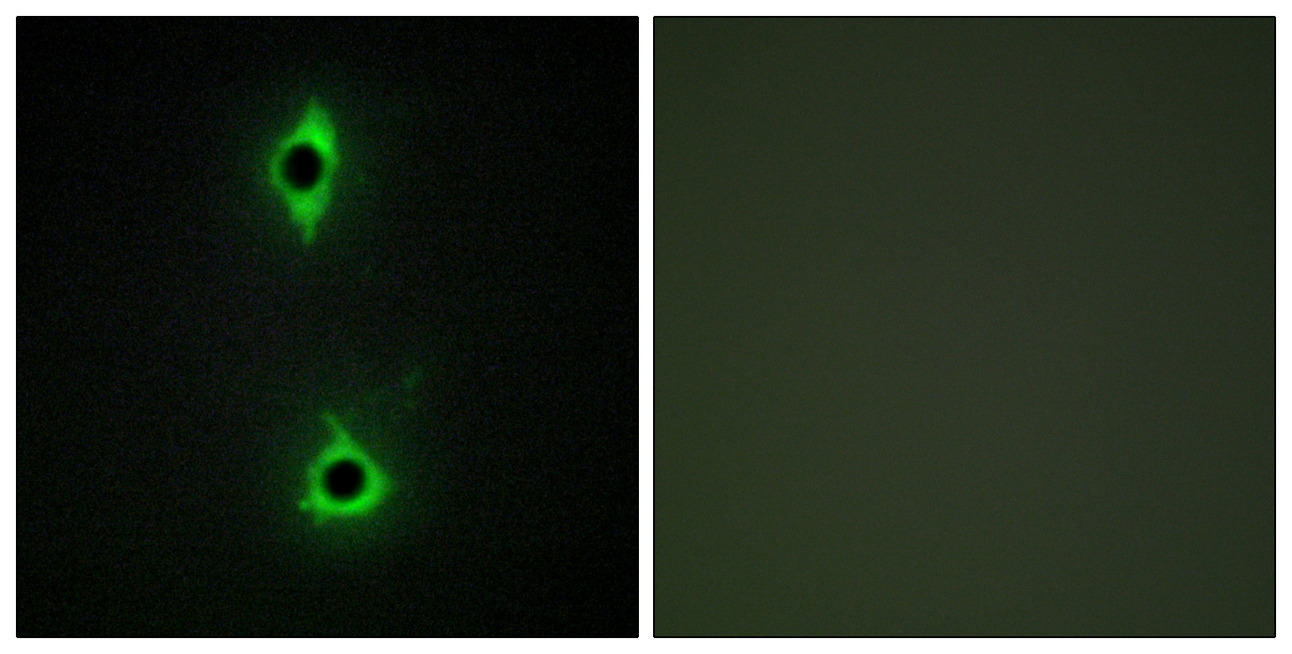
- Immunofluorescence analysis of COS7 cells, using PIP5K Antibody. The picture on the right is blocked with the synthesized peptide.
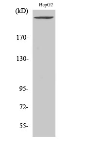
- Western blot analysis of lysates from HepG2 cells, using PIP5K Antibody. The lane on the right is blocked with the synthesized peptide.
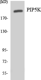
- Western blot analysis of the lysates from Jurkat cells using PIP5K antibody.

- Immunohistochemical analysis of paraffin-embedded human uterus. 1, Antibody was diluted at 1:200(4° overnight). 2, Tris-EDTA,pH9.0 was used for antigen retrieval. 3,Secondary antibody was diluted at 1:200(room temperature, 45min).