PCAF Polyclonal Antibody
- Catalog No.:YT3615
- Applications:WB;IHC;IF;ELISA
- Reactivity:Human;Mouse;Rat
- Target:
- PCAF
- Fields:
- >>Viral life cycle - HIV-1;>>Notch signaling pathway;>>Thyroid hormone signaling pathway;>>Human T-cell leukemia virus 1 infection;>>Viral carcinogenesis
- Gene Name:
- KAT2B
- Protein Name:
- Histone acetyltransferase KAT2B
- Human Gene Id:
- 8850
- Human Swiss Prot No:
- Q92831
- Mouse Gene Id:
- 18519
- Mouse Swiss Prot No:
- Q9JHD1
- Immunogen:
- The antiserum was produced against synthesized peptide derived from human p300/CBP. AA range:783-832
- Specificity:
- PCAF Polyclonal Antibody detects endogenous levels of PCAF protein.
- Formulation:
- Liquid in PBS containing 50% glycerol, 0.5% BSA and 0.02% sodium azide.
- Source:
- Polyclonal, Rabbit,IgG
- Dilution:
- WB 1:500 - 1:2000. IHC 1:100 - 1:300. ELISA: 1:10000.. IF 1:50-200
- Purification:
- The antibody was affinity-purified from rabbit antiserum by affinity-chromatography using epitope-specific immunogen.
- Concentration:
- 1 mg/ml
- Storage Stability:
- -15°C to -25°C/1 year(Do not lower than -25°C)
- Other Name:
- KAT2B;PCAF;Histone acetyltransferase KAT2B;Histone acetyltransferase PCAF;Histone acetylase PCAF;Lysine acetyltransferase 2B;P300/CBP-associated factor;P/CAF
- Observed Band(KD):
- 93kD
- Background:
- CBP and p300 are large nuclear proteins that bind to many sequence-specific factors involved in cell growth and/or differentiation, including c-jun and the adenoviral oncoprotein E1A. The protein encoded by this gene associates with p300/CBP. It has in vitro and in vivo binding activity with CBP and p300, and competes with E1A for binding sites in p300/CBP. It has histone acetyl transferase activity with core histones and nucleosome core particles, indicating that this protein plays a direct role in transcriptional regulation. [provided by RefSeq, Jul 2008],
- Function:
- chromatin organization, chromatin remodeling, transcription, regulation of transcription, DNA-dependent, protein amino acid acetylation, N-terminal protein amino acid acetylation, cell cycle, cell cycle arrest, negative regulation of cell proliferation, response to endogenous stimulus, response to hormone stimulus, response to organic substance,chromatin modification, covalent chromatin modification, histone modification, histone acetylation, N-terminal peptidyl-lysine acetylation, peptidyl-lysine modification, peptidyl-lysine acetylation, cell cycle process, N-terminal protein amino acid modification, response to insulin stimulus, cellular response to insulin stimulus, cellular response to hormone stimulus, regulation of cell proliferation, response to peptide hormone stimulus, protein amino acid acylation,regulation of transcription, regulation of RNA metabolic process, chromosome orga
- Subcellular Location:
- Nucleus . Cytoplasm, cytoskeleton, microtubule organizing center, centrosome . Cytoplasm . Mainly localizes to the nucleus. Also localizes to centrosomes in late G1 and around the G1/S transition, coinciding with the onset of centriole formation. Subcellular location may vary depending upon cell differentiation state. Cytoplasmic at the very stages of keratinocyte differentiation, becomes nuclear at later differentiation stages. Cytoplasmic in basal epithelial cells (undifferentiated cells) and nuclear in parabasal cells (differentiated cells) (PubMed:20940255). .
- Expression:
- Ubiquitously expressed but most abundant in heart and skeletal muscle. Also expressed in the skin, in keratinocytes (at protein level) (PubMed:20940255).
- June 19-2018
- WESTERN IMMUNOBLOTTING PROTOCOL
- June 19-2018
- IMMUNOHISTOCHEMISTRY-PARAFFIN PROTOCOL
- June 19-2018
- IMMUNOFLUORESCENCE PROTOCOL
- September 08-2020
- FLOW-CYTOMEYRT-PROTOCOL
- May 20-2022
- Cell-Based ELISA│解您多样本WB检测之困扰
- July 13-2018
- CELL-BASED-ELISA-PROTOCOL-FOR-ACETYL-PROTEIN
- July 13-2018
- CELL-BASED-ELISA-PROTOCOL-FOR-PHOSPHO-PROTEIN
- July 13-2018
- Antibody-FAQs
- Products Images
- Western Blot analysis of various cells using PCAF Polyclonal Antibody cells nucleus extracted by Minute TM Cytoplasmic and Nuclear Fractionation kit (SC-003,Inventbiotech,MN,USA).
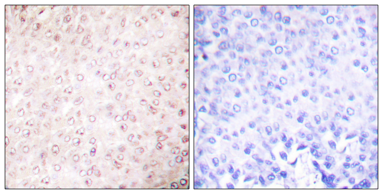
- Immunohistochemistry analysis of paraffin-embedded human breast carcinoma tissue, using p300/CBP Antibody. The picture on the right is blocked with the synthesized peptide.
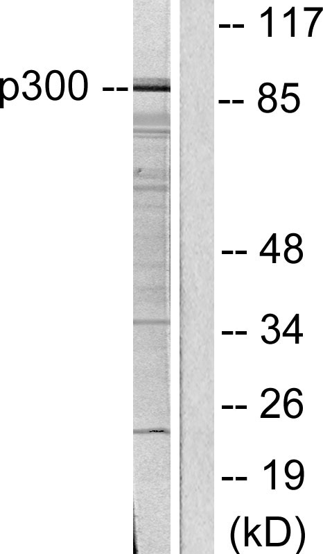
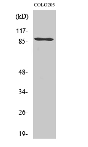
- Western blot analysis of lysates from COLO205 cells, using p300/CBP Antibody. The lane on the right is blocked with the synthesized peptide.
.jpg)
