Lamin A/C (phospho Ser392) (PTR1133) mouse mAb
- Catalog No.:YP1876
- Applications:WB;IF;ELISA
- Reactivity:Human;Mouse;Rat;Monkey;
- Target:
- Lamin A/C (phospho Ser392)
- Gene Name:
- LMNA LMN1
- Protein Name:
- Prelamin-A/C [Cleaved into: Lamin-A/C (70 kDa lamin) (Renal carcinoma antigen NY-REN-32)]
- Human Gene Id:
- 4000
- Human Swiss Prot No:
- P02545
- Mouse Gene Id:
- 16905
- Mouse Swiss Prot No:
- P48678
- Immunogen:
- Synthesized peptide derived from human protein. AA range: 350-450
- Specificity:
- This antibody detects endogenous levels of Lamin A/C (phospho Ser392) protein.
- Formulation:
- PBS, 50% glycerol, 0.05% Proclin 300, 0.05%BSA
- Source:
- Mouse, Monoclonal/IgG1, kappa
- Dilution:
- WB 1:500-2000. IF 1:100-500. ELISA 1:1000-5000
- Purification:
- Protein G
- Concentration:
- 1 mg/ml
- Storage Stability:
- -15°C to -25°C/1 year(Do not lower than -25°C)
- Other Name:
- Prelamin-A/C [Cleaved into: Lamin-A/C (70 kDa lamin) (Renal carcinoma antigen NY-REN-32)]
- Molecular Weight(Da):
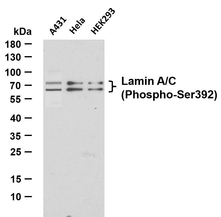
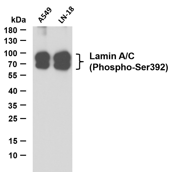
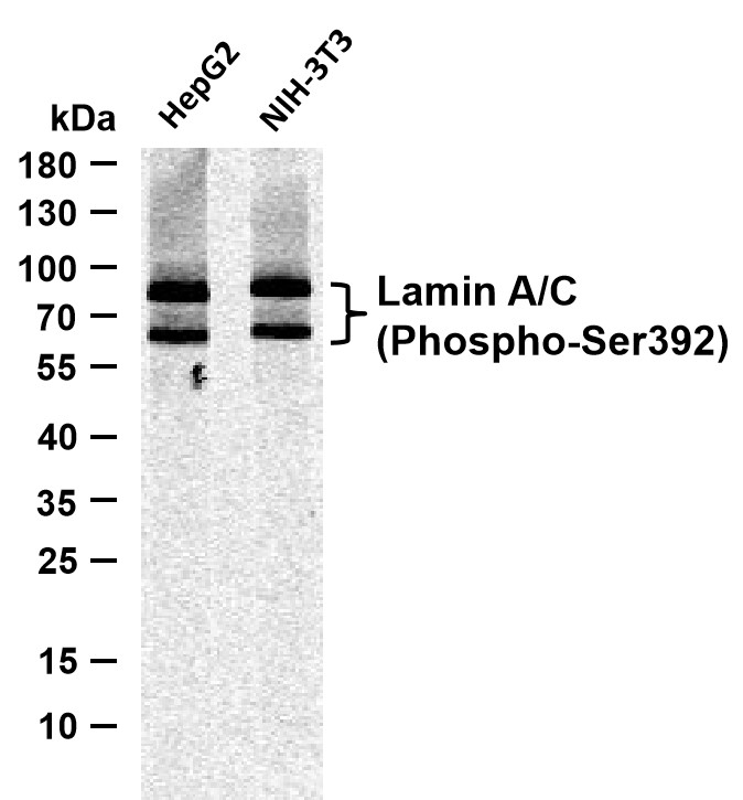
- 63kD,74kD
- Observed Band(KD):
- 63kD,74kD
- Background:
- lamin A/C(LMNA) Homo sapiens The nuclear lamina consists of a two-dimensional matrix of proteins located next to the inner nuclear membrane. The lamin family of proteins make up the matrix and are highly conserved in evolution. During mitosis, the lamina matrix is reversibly disassembled as the lamin proteins are phosphorylated. Lamin proteins are thought to be involved in nuclear stability, chromatin structure and gene expression. Vertebrate lamins consist of two types, A and B. Alternative splicing results in multiple transcript variants. Mutations in this gene lead to several diseases: Emery-Dreifuss muscular dystrophy, familial partial lipodystrophy, limb girdle muscular dystrophy, dilated cardiomyopathy, Charcot-Marie-Tooth disease, and Hutchinson-Gilford progeria syndrome. [provided by RefSeq, Apr 2012],
- Function:
- Lamins are components of the nuclear lamina, a fibrous layer on the nucleoplasmic side of the inner nuclear membrane, which is thought to provide a framework for the nuclear envelope and may also interact with chromatin. Lamin A and C are present in equal amounts in the lamina of mammals. Recruited by DNA repair proteins XRCC4 and IFFO1 to the DNA double-strand breaks (DSBs) to prevent chromosome translocation by immobilizing broken DNA ends . Plays an important role in nuclear assembly, chromatin organization, nuclear membrane and telomere dynamics. Required for normal development of peripheral nervous system and skeletal muscle and for muscle satellite cell proliferation . Required for osteoblastogenesis and bone formation . Also prevents fat infiltration of muscle and bone marrow, helping to maintain the volume and strength of skeletal muscle and bone . Required for cardiac homeostas
- Subcellular Location:
- 0
- Expression:
- In the arteries, prelamin-A/C accumulation is not observed in young healthy vessels but is prevalent in medial vascular smooth muscle cells (VSMCs) from aged individuals and in atherosclerotic lesions, where it often colocalizes with senescent and degenerate VSMCs. Prelamin-A/C expression increases with age and disease. In normal aging, the accumulation of prelamin-A/C is caused in part by the down-regulation of ZMPSTE24/FACE1 in response to oxidative stress.
- June 19-2018
- WESTERN IMMUNOBLOTTING PROTOCOL
- June 19-2018
- IMMUNOHISTOCHEMISTRY-PARAFFIN PROTOCOL
- June 19-2018
- IMMUNOFLUORESCENCE PROTOCOL
- September 08-2020
- FLOW-CYTOMEYRT-PROTOCOL
- May 20-2022
- Cell-Based ELISA│解您多样本WB检测之困扰
- July 13-2018
- CELL-BASED-ELISA-PROTOCOL-FOR-ACETYL-PROTEIN
- July 13-2018
- CELL-BASED-ELISA-PROTOCOL-FOR-PHOSPHO-PROTEIN
- July 13-2018
- Antibody-FAQs
- Products Images

- Various whole cell lysates were separated by 10% SDS-PAGE, and the membrane was blotted with anti-Lamin A/C (Phospho-Ser392) (PTR1133) antibody. The HRP-conjugated Goat anti-Mouse IgG(H + L) antibody was used to detect the antibody. Lane 1: A431 Lane 2: Hela Lane 3: HEK293

- Various whole cell lysates were separated by 10% SDS-PAGE, and the membrane was blotted with anti-Lamin A/C (Phospho-Ser392) (PTR1133) antibody. The HRP-conjugated Goat anti-Mouse IgG(H + L) antibody was used to detect the antibody. Lane 1: A549 Lane 2: LN18

- Various whole cell lysates were separated by 10% SDS-PAGE, and the membrane was blotted with anti-Lamin A/C (Phospho-Ser392) (PTR1133) antibody. The HRP-conjugated Goat anti-Mouse IgG(H + L) antibody was used to detect the antibody. Lane 1: HepG2 Lane 2: NIH-3T3

