Stat3 (phospho Tyr705) Polyclonal Antibody
- Catalog No.:YP0251
- Applications:IF;WB;IHC;IP;ELISA
- Reactivity:Human;Mouse;Rat;Pig(Test by out customer)
- Target:
- Stat3
- Fields:
- >>EGFR tyrosine kinase inhibitor resistance;>>Chemokine signaling pathway;>>HIF-1 signaling pathway;>>FoxO signaling pathway;>>Necroptosis;>>Signaling pathways regulating pluripotency of stem cells;>>JAK-STAT signaling pathway;>>Th17 cell differentiation;>>Prolactin signaling pathway;>>Adipocytokine signaling pathway;>>Insulin resistance;>>AGE-RAGE signaling pathway in diabetic complications;>>Growth hormone synthesis, secretion and action;>>Toxoplasmosis;>>Hepatitis C;>>Hepatitis B;>>Measles;>>Human cytomegalovirus infection;>>Kaposi sarcoma-associated herpesvirus infection;>>Epstein-Barr virus infection;>>Coronavirus disease - COVID-19;>>Pathways in cancer;>>Viral carcinogenesis;>>Proteoglycans in cancer;>>MicroRNAs in cancer;>>Chemical carcinogenesis - receptor activation;>>Pancreatic cancer;>>Acute myeloid leukemia;>>Non-small cell lung cancer;>>PD-L1 expression and PD-1 checkpoint pathway in cancer;>>Inflammatory bowel disease;>>Lipid and atherosclerosis
- Gene Name:
- STAT3
- Protein Name:
- Signal transducer and activator of transcription 3
- Human Gene Id:
- 6774
- Human Swiss Prot No:
- P40763
- Mouse Gene Id:
- 20848
- Mouse Swiss Prot No:
- P42227
- Rat Gene Id:
- 25125
- Rat Swiss Prot No:
- P52631
- Immunogen:
- The antiserum was produced against synthesized peptide derived from human STAT3 around the phosphorylation site of Tyr705. AA range:672-721
- Specificity:
- Phospho-Stat3 (Y705) Polyclonal Antibody detects endogenous levels of Stat3 protein only when phosphorylated at Y705.
- Formulation:
- Liquid in PBS containing 50% glycerol, 0.5% BSA and 0.02% sodium azide.
- Source:
- Polyclonal, Rabbit,IgG
- Dilution:
- IF 1:50-200 WB 1:500 - 1:2000. IHC 1:100 - 1:300. Immunoprecipitation: 2-5 ug:mg lysate. ELISA: 1:20000. Not yet tested in other applications.
- Purification:
- The antibody was affinity-purified from rabbit antiserum by affinity-chromatography using epitope-specific immunogen.
- Concentration:
- 1 mg/ml
- Storage Stability:
- -15°C to -25°C/1 year(Do not lower than -25°C)
- Other Name:
- STAT3;APRF;Signal transducer and activator of transcription 3;Acute-phase response factor
- Observed Band(KD):
- 88kD
- Background:
- The protein encoded by this gene is a member of the STAT protein family. In response to cytokines and growth factors, STAT family members are phosphorylated by the receptor associated kinases, and then form homo- or heterodimers that translocate to the cell nucleus where they act as transcription activators. This protein is activated through phosphorylation in response to various cytokines and growth factors including IFNs, EGF, IL5, IL6, HGF, LIF and BMP2. This protein mediates the expression of a variety of genes in response to cell stimuli, and thus plays a key role in many cellular processes such as cell growth and apoptosis. The small GTPase Rac1 has been shown to bind and regulate the activity of this protein. PIAS3 protein is a specific inhibitor of this protein. Mutations in this gene are associated with infantile-onset multisystem autoimmune disease and hyper
- Function:
- disease:Defects in STAT3 are the cause of hyperimmunoglobulin E recurrent infection syndrome autosomal dominant (AD-HIES) [MIM:147060]; also known as hyper-IgE syndrome or Job syndrome. AD-HIES is a rare disorder of immunity and connective tissue characterized by immunodeficiency, chronic eczema, recurrent Staphylococcal infections, increased serum IgE, eosinophilia, distinctive coarse facial appearance, abnormal dentition, hyperextensibility of the joints, and bone fractures.,function:Transcription factor that binds to the interleukin-6 (IL-6)-responsive elements identified in the promoters of various acute-phase protein genes. Activated by IL31 through IL31RA.,miscellaneous:Involved in the gp130-mediated signaling pathway.,online information:STAT3 entry,online information:STAT3 mutation db,PTM:Tyrosine phosphorylated in response to IL-6, IL-11, CNTF, LIF, CSF-1, EGF, PDGF, IFN-alpha an
- Subcellular Location:
- Cytoplasm . Nucleus . Shuttles between the nucleus and the cytoplasm. Translocated into the nucleus upon tyrosine phosphorylation and dimerization, in response to signaling by activated FGFR1, FGFR2, FGFR3 or FGFR4. Constitutive nuclear presence is independent of tyrosine phosphorylation. Predominantly present in the cytoplasm without stimuli. Upon leukemia inhibitory factor (LIF) stimulation, accumulates in the nucleus. The complex composed of BART and ARL2 plays an important role in the nuclear translocation and retention of STAT3. Identified in a complex with LYN and PAG1.
- Expression:
- Heart, brain, placenta, lung, liver, skeletal muscle, kidney and pancreas. Expressed in naive CD4(+) T cells as well as T-helper Th17, Th1 and Th2 cells (PubMed:31899195).
Afatinib Reverses EMT via Inhibiting CD44-Stat3 Axis to Promote Radiosensitivity in Nasopharyngeal Carcinoma Pharmaceuticals Huichao Huang, Fangling Huang, Xujun Liang, Ying Fu, Zhe Cheng, Yan Huang, Zhuchu Chen, Yankun Duan, Yongheng Chen WB Human 5-8F cell, HNE2 cell
Suppression of the hyaluronic acid pathway induces M1 macrophages polarization via STAT1 in glioblastoma. Cell Death Discovery2022 Apr;8(1):1-13. Human U937 monocytes,U251 cell,LN229 cell
Salmonella effector SpvB aggravates dysregulation of systemic iron metabolism via modulating the hepcidin−ferroportin axis. Gut Microbes Gut Microbes. 2021;13(1):1-18 WB Mouse Liver
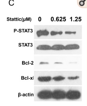
Sabutoclax, pan-active BCL-2 protein family antagonist, overcomes drug resistance and eliminates cancer stem cells in breast cancer. CANCER LETTERS 2018 Feb 27 WB,IHC Human breast cancer tissue MCF-7 cell,CALDOX cell,MCF-7/A02 cell,Cal51 cell
HIF‐1α facilitates osteocyte‐mediated osteoclastogenesis by activating JAK2/STAT3 pathway in vitro. JOURNAL OF CELLULAR PHYSIOLOGY 2019 Apr 29 WB Mouse 1:500 MLO-Y4 cell
Integrative proteomics and immunochemistry analysis of the factors in the necrosis and repair in acetaminophen‐induced acute liver injury in mice. JOURNAL OF CELLULAR PHYSIOLOGY J Cell Physiol. 2019 May;234(5):6561-6581 WB Mouse 1:100 liver
Lapatinib‑induced inhibition of ovarian function is counteracted by the STAT3 pathway both in vivo and in vitro. ONCOLOGY REPORTS Oncol Rep. 2020 Sep;44(3):1127-1135 IHC Mouse 1:50 Ovarian tissue Oocytes
-Induced Injury in Induced Pluripotent Stem Cell-Derived Neural Stem Cells. NEUROCHEMICAL RESEARCH Neurochem Res. 2015 Jun;40(6):1133-1143 WB Mouse 1:1000 iPSC-derived NSCs
Shu, Tao, et al. "Protective effects and mechanisms of salvianolic acid B against H2O2-Induced injury in induced pluripotent stem cell-derived neural stem cells." Neurochemical research 40.6 (2015): 1133-1143.
Anti-tumor effects of novel alkannin derivatives with potent selectivity on comprehensive analysis. Hai-min Lei WB Human A549 cell
G3BP1 Interact with JAK2 mRNA to Promote the Malignant Progression of Nasopharyngeal Carcinoma via Activating JAK2/STAT3 Signaling Pathway International Journal of Biological Sciences Zhan Yuting IHC Human 1:250 nasopharyngeal epithelial tissue
Exosomes Derived from Adipose Mesenchymal Stem Cells Promote Regeneration of Injured Liver in Minipigs INTERNATIONAL JOURNAL OF MOLECULAR SCIENCES Yue Wang WB Pig 1:1000 liver tissue
Gastrodin against oxidative stress-inflammation crosstalk via inhibiting mtDNA/TLR9 and JAK2/STAT3 signaling to ameliorate ischemic stroke injury INTERNATIONAL IMMUNOPHARMACOLOGY Menglian Zhang WB Rat 1:1000 brain tissue brain microvascular endothelial cell (BMEC)
- June 19-2018
- WESTERN IMMUNOBLOTTING PROTOCOL
- June 19-2018
- IMMUNOHISTOCHEMISTRY-PARAFFIN PROTOCOL
- June 19-2018
- IMMUNOFLUORESCENCE PROTOCOL
- September 08-2020
- FLOW-CYTOMEYRT-PROTOCOL
- May 20-2022
- Cell-Based ELISA│解您多样本WB检测之困扰
- July 13-2018
- CELL-BASED-ELISA-PROTOCOL-FOR-ACETYL-PROTEIN
- July 13-2018
- CELL-BASED-ELISA-PROTOCOL-FOR-PHOSPHO-PROTEIN
- July 13-2018
- Antibody-FAQs
- Products Images
- Liu, Yanmei, et al. "Cancer Stem Cells are Regulated by STAT3 Signalling in Wilms Tumour." Journal of Cancer 9.8 (2018): 1486.
-if-104.jpg)
- Immunofluorescence analysis of mouse-spleen tissue. 1,Stat3 (phospho Tyr705) Polyclonal Antibody(red) was diluted at 1:200(4°C,overnight). 2, Cy3 labled Secondary antibody was diluted at 1:300(room temperature, 50min).3, Picture B: DAPI(blue) 10min. Picture A:Target. Picture B: DAPI. Picture C: merge of A+B
-if-105.jpg)
- Immunofluorescence analysis of mouse-spleen tissue. 1,Stat3 (phospho Tyr705) Polyclonal Antibody(red) was diluted at 1:200(4°C,overnight). 2, Cy3 labled Secondary antibody was diluted at 1:300(room temperature, 50min).3, Picture B: DAPI(blue) 10min. Picture A:Target. Picture B: DAPI. Picture C: merge of A+B
poly-ihc-human-lung-cancer.jpg)
- Immunohistochemical analysis of paraffin-embedded Human-lung-cancer tissue. 1,Stat3 (phospho Tyr705) Polyclonal Antibody was diluted at 1:200(4°C,overnight). 2, Sodium citrate pH 6.0 was used for antibody retrieval(>98°C,20min). 3,Secondary antibody was diluted at 1:200(room tempeRature, 30min). Negative control was used by secondary antibody only.
poly-ihc-rat-lung.jpg)
- Immunohistochemical analysis of paraffin-embedded Rat-lung tissue. 1,Stat3 (phospho Tyr705) Polyclonal Antibody was diluted at 1:200(4°C,overnight). 2, Sodium citrate pH 6.0 was used for antibody retrieval(>98°C,20min). 3,Secondary antibody was diluted at 1:200(room tempeRature, 30min). Negative control was used by secondary antibody only.
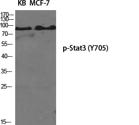
- Western Blot analysis of various cells using Phospho-Stat3 (Y705) Polyclonal Antibody diluted at 1:2000
.jpg)
- Western Blot analysis of KB cells using Phospho-Stat3 (Y705) Polyclonal Antibody diluted at 1:2000
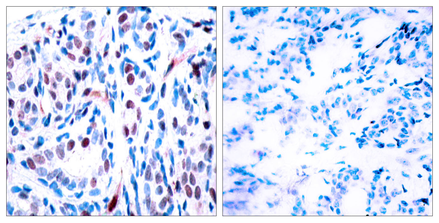
- Immunohistochemistry analysis of paraffin-embedded human breast carcinoma, using STAT3 (Phospho-Tyr705) Antibody. The picture on the right is blocked with the phospho peptide.

- Western blot analysis of lysates from HeLa cells, using STAT3 (Phospho-Tyr705) Antibody. The lane on the right is blocked with the phospho peptide.

