pVHL (ABT-PVHL) mouse mAb
- Catalog No.:YM6215
- Applications:IHC;IF;ELISA
- Reactivity:Human;
- Target:
- VHL
- Fields:
- >>HIF-1 signaling pathway;>>Ubiquitin mediated proteolysis;>>Pathways in cancer;>>Renal cell carcinoma
- Gene Name:
- VHL
- Protein Name:
- Von Hippel-Lindau disease tumor suppressor (Protein G7) (pVHL)
- Human Gene Id:
- 7428
- Human Swiss Prot No:
- P40337
- Immunogen:
- Synthesized peptide derived from human pVHL AA range: 150-213
- Specificity:
- This antibody detects endogenous levels of pVHL protein.
- Formulation:
- PBS, 50% glycerol, 0.05% Proclin 300, 0.05%BSA
- Source:
- Mouse, Monoclonal/IgG2b, kappa
- Dilution:
- IHC 1:50-200. IF 1:50-200. ELISA 1:500-5000
- Purification:
- The antibody was affinity-purified from ascites by affinity-chromatography using specific immunogen.
- Storage Stability:
- -15°C to -25°C/1 year(Do not lower than -25°C)
- Molecular Weight(Da):
- 24kD,19kD
- Observed Band(KD):
- 17kD
- Background:
- von Hippel-Lindau tumor suppressor(VHL) Homo sapiens Von Hippel-Lindau syndrome (VHL) is a dominantly inherited familial cancer syndrome predisposing to a variety of malignant and benign tumors. A germline mutation of this gene is the basis of familial inheritance of VHL syndrome. The protein encoded by this gene is a component of the protein complex that includes elongin B, elongin C, and cullin-2, and possesses ubiquitin ligase E3 activity. This protein is involved in the ubiquitination and degradation of hypoxia-inducible-factor (HIF), which is a transcription factor that plays a central role in the regulation of gene expression by oxygen. RNA polymerase II subunit POLR2G/RPB7 is also reported to be a target of this protein. Alternatively spliced transcript variants encoding distinct isoforms have been observed. [provided by RefSeq, Jul 2008],
- Function:
- disease:Defects in VHL are a cause of pheochromocytoma [MIM:171300]. The pheochromocytomas are catecholamine-producing, chromaffin tumors that arise in the adrenal medulla in 90% of cases. In the remaining 10% of cases, they develop in extra-adrenal sympathetic ganglia and may be referred to as "paraganglioma." Pheochromocytoma usually presents with hypertension. Approximately 10% of pheochromocytoma is hereditary. The genetic basis for most cases of non-syndromic familial pheochromocytoma is unknown.,disease:Defects in VHL are a cause of renal cell carcinoma type 1 (RCC1) [MIM:144700]; also called hypernephroma or adenocarcinoma of kidney. Familial renal cell carcinoma syndromes form a group of diseases characterized by a predisposition to development of renal cell carcinomas (RCCs) with various histological subtypes.,disease:Defects in VHL are the cause of erythrocytosis familial type
- Subcellular Location:
- Cytoplasmic
- Expression:
- Expressed in the adult and fetal brain and kidney.
- June 19-2018
- WESTERN IMMUNOBLOTTING PROTOCOL
- June 19-2018
- IMMUNOHISTOCHEMISTRY-PARAFFIN PROTOCOL
- June 19-2018
- IMMUNOFLUORESCENCE PROTOCOL
- September 08-2020
- FLOW-CYTOMEYRT-PROTOCOL
- May 20-2022
- Cell-Based ELISA│解您多样本WB检测之困扰
- July 13-2018
- CELL-BASED-ELISA-PROTOCOL-FOR-ACETYL-PROTEIN
- July 13-2018
- CELL-BASED-ELISA-PROTOCOL-FOR-PHOSPHO-PROTEIN
- July 13-2018
- Antibody-FAQs
- Products Images
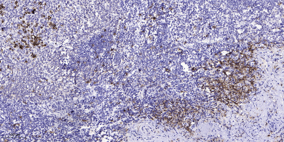
- Human Kidney tissue was stained with Anti-pVHL (ABT-PVHL) Antibody

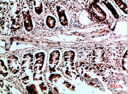
- Human liver tissue was stained with Anti-pVHL (ABT-PVHL) Antibody

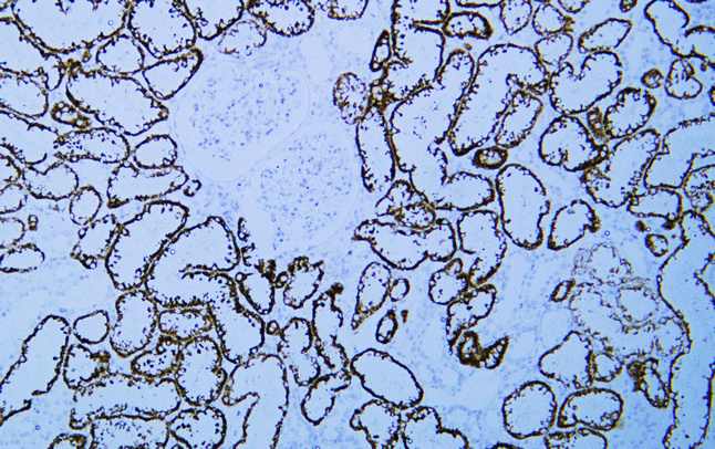
- Human pancreas tissue was stained with Anti-pVHL (ABT-PVHL) Antibody
.jpg)
- Immunohistochemical analysis of paraffin-embedded kidney. 1, Antibody was diluted at 1:200(4° overnight). 2, Tris-EDTA,pH8.0 was used for antigen retrieval. 3,Secondary antibody was diluted at 1:200(room temperature, 30min).
.jpg)
- Immunohistochemical analysis of paraffin-embedded kidney. 1, Antibody was diluted at 1:200(4° overnight). 2, Tris-EDTA,pH8.0 was used for antigen retrieval. 3,Secondary antibody was diluted at 1:200(room temperature, 30min).
.jpg)
- Immunohistochemical analysis of paraffin-embedded kidney. 1, Antibody was diluted at 1:200(4° overnight). 2, Tris-EDTA,pH8.0 was used for antigen retrieval. 3,Secondary antibody was diluted at 1:200(room temperature, 30min).
.jpg)
- Immunohistochemical analysis of paraffin-embedded kidney. 1, Antibody was diluted at 1:200(4° overnight). 2, Tris-EDTA,pH8.0 was used for antigen retrieval. 3,Secondary antibody was diluted at 1:200(room temperature, 30min).
.jpg)
- Immunohistochemical analysis of paraffin-embedded kidney. 1, Antibody was diluted at 1:200(4° overnight). 2, Tris-EDTA,pH8.0 was used for antigen retrieval. 3,Secondary antibody was diluted at 1:200(room temperature, 30min).

- Immunohistochemical analysis of paraffin-embedded Liver. 1, Antibody was diluted at 1:200(4° overnight). 2, Tris-EDTA,pH8.0 was used for antigen retrieval. 3,Secondary antibody was diluted at 1:200(room temperature, 30min).