R-Cadherin (PT2127) mouse mAb
- Catalog No.:YM4300
- Applications:WB;IF;ELISA
- Reactivity:Human;Mouse;Rat;
- Target:
- CDH4
- Gene Name:
- CDH4
- Protein Name:
- Cadherin-4 (Retinal cadherin) (R-CAD) (R-cadherin)
- Human Gene Id:
- 1002
- Human Swiss Prot No:
- P55283
- Mouse Gene Id:
- 12561
- Mouse Swiss Prot No:
- P39038
- Rat Swiss Prot No:
- Q63149
- Immunogen:
- Synthesized peptide derived from human R-Cadherin. AA range: 150-250
- Specificity:
- This antibody detects endogenous levels of R-Cadherin protein.
- Formulation:
- PBS, 50% glycerol, 0.05% Proclin 300, 0.05%BSA
- Source:
- Mouse, Monoclonal/IgG1, kappa
- Dilution:
- WB 1:500-2000. IF 1:100-500. ELISA 1:1000-5000
- Purification:
- Protein G
- Concentration:
- 1 mg/ml
- Storage Stability:
- -15°C to -25°C/1 year(Do not lower than -25°C)
- Other Name:
- Cadherin-4 (Retinal cadherin) (R-CAD) (R-cadherin)
- Molecular Weight(Da):
- 102kD
- Observed Band(KD):
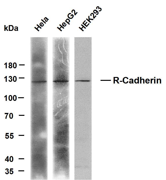
- 120kD
- Background:
- cadherin 4(CDH4) Homo sapiens This gene is a classical cadherin from the cadherin superfamily. The encoded protein is a calcium-dependent cell-cell adhesion glycoprotein comprised of five extracellular cadherin repeats, a transmembrane region and a highly conserved cytoplasmic tail. Based on studies in chicken and mouse, this cadherin is thought to play an important role during brain segmentation and neuronal outgrowth. In addition, a role in kidney and muscle development is indicated. Of particular interest are studies showing stable cis-heterodimers of cadherins 2 and 4 in cotransfected cell lines. Previously thought to interact in an exclusively homophilic manner, this is the first evidence of cadherin heterodimerization. Three transcript variants encoding different isoforms have been found for this gene. [provided by RefSeq, Nov 2011],
- Function:
- Cadherins are calcium-dependent cell adhesion proteins. They preferentially interact with themselves in a homophilic manner in connecting cells; cadherins may thus contribute to the sorting of heterogeneous cell types. May play an important role in retinal development.
- Subcellular Location:
- Membranous
- Expression:
- Expressed mainly in brain but also found in other tissues.
- June 19-2018
- WESTERN IMMUNOBLOTTING PROTOCOL
- June 19-2018
- IMMUNOHISTOCHEMISTRY-PARAFFIN PROTOCOL
- June 19-2018
- IMMUNOFLUORESCENCE PROTOCOL
- September 08-2020
- FLOW-CYTOMEYRT-PROTOCOL
- May 20-2022
- Cell-Based ELISA│解您多样本WB检测之困扰
- July 13-2018
- CELL-BASED-ELISA-PROTOCOL-FOR-ACETYL-PROTEIN
- July 13-2018
- CELL-BASED-ELISA-PROTOCOL-FOR-PHOSPHO-PROTEIN
- July 13-2018
- Antibody-FAQs
- Products Images

- Various whole cell lysates were separated by 8% SDS-PAGE, and the membrane was blotted with anti-R-Cadherin (PT2127) antibody. The HRP-conjugated anti-Mouse IgG antibody was used to detect the antibody. Lane 1: Hela Lane 2: HepG2 Lane 3: HEK293

