PDGFRα mouse Monoclonal Antibody(7A3)
- Catalog No.:YM3687
- Applications:IF;IHC
- Reactivity:Human;Rat;Mouse
- Target:
- PDGF Receptor α
- Fields:
- >>EGFR tyrosine kinase inhibitor resistance;>>MAPK signaling pathway;>>Ras signaling pathway;>>Rap1 signaling pathway;>>Calcium signaling pathway;>>Phospholipase D signaling pathway;>>Endocytosis;>>PI3K-Akt signaling pathway;>>Focal adhesion;>>Gap junction;>>JAK-STAT signaling pathway;>>Regulation of actin cytoskeleton;>>Human cytomegalovirus infection;>>Pathways in cancer;>>MicroRNAs in cancer;>>Glioma;>>Prostate cancer;>>Melanoma;>>Central carbon metabolism in cancer;>>Choline metabolism in cancer
- Gene Name:
- PDGFRA
- Protein Name:
- PDGFRA
- Human Gene Id:
- 5156
- Human Swiss Prot No:
- P16234
- Mouse Swiss Prot No:
- P26618
- Rat Swiss Prot No:
- P20786
- Immunogen:
- Synthetic Peptide of PDGFRα at AA range of 1010-1090
- Specificity:
- PDGFRα protein detects endogenous levels of PDGFRA
- Formulation:
- Liquid in PBS containing 50% glycerol, 0.5% BSA and 0.02% sodium azide.
- Source:
- Monoclonal, Mouse
- Dilution:
- IF 1:50-200 IHC 1:100-200
- Purification:
- The antibody was affinity-purified from mouse ascites by affinity-chromatography using specific immunogen.
- Concentration:
- 1 mg/ml
- Storage Stability:
- -15°C to -25°C/1 year(Do not lower than -25°C)
- Other Name:
- PDGFRA
- Observed Band(KD):
- 180kD
- Background:
- This gene encodes a cell surface tyrosine kinase receptor for members of the platelet-derived growth factor family. These growth factors are mitogens for cells of mesenchymal origin. The identity of the growth factor bound to a receptor monomer determines whether the functional receptor is a homodimer or a heterodimer, composed of both platelet-derived growth factor receptor alpha and beta polypeptides. Studies suggest that this gene plays a role in organ development, wound healing, and tumor progression. Mutations in this gene have been associated with idiopathic hypereosinophilic syndrome, somatic and familial gastrointestinal stromal tumors, and a variety of other cancers. [provided by RefSeq, Mar 2012],
- Function:
- catalytic activity:ATP + a [protein]-L-tyrosine = ADP + a [protein]-L-tyrosine phosphate.,disease:A fusion of PDGFRA and FIP1L1 (FIP1L1-PDGFRA), due to an interstitial chromosomal deletion, is the cause of some cases of hypereosinophilic syndrome (HES) [MIM:607685]. HES is a rare hematologic disorder characterized by sustained overproduction of eosinophils in the bone marrow, eosinophilia, tissue infiltration and organ damage.,function:Receptor that binds both PDGFA and PDGFB and has a tyrosine-protein kinase activity.,similarity:Belongs to the protein kinase superfamily. Tyr protein kinase family. CSF-1/PDGF receptor subfamily.,similarity:Contains 1 protein kinase domain.,similarity:Contains 5 Ig-like C2-type (immunoglobulin-like) domains.,subunit:Homodimer, and heterodimer with PDGFRB. Interacts with the SH2 domain of SHB via phosphorylated Tyr-720 (By similarity). Interacts with the S
- Subcellular Location:
- Cell membrane ; Single-pass type I membrane protein . Cell projection, cilium . Golgi apparatus .
- Expression:
- Detected in platelets (at protein level). Widely expressed. Detected in brain, fibroblasts, smooth muscle, heart, and embryo. Expressed in primary and metastatic colon tumors and in normal colon tissue.
- June 19-2018
- WESTERN IMMUNOBLOTTING PROTOCOL
- June 19-2018
- IMMUNOHISTOCHEMISTRY-PARAFFIN PROTOCOL
- June 19-2018
- IMMUNOFLUORESCENCE PROTOCOL
- September 08-2020
- FLOW-CYTOMEYRT-PROTOCOL
- May 20-2022
- Cell-Based ELISA│解您多样本WB检测之困扰
- July 13-2018
- CELL-BASED-ELISA-PROTOCOL-FOR-ACETYL-PROTEIN
- July 13-2018
- CELL-BASED-ELISA-PROTOCOL-FOR-PHOSPHO-PROTEIN
- July 13-2018
- Antibody-FAQs
- Products Images

- Immunofluorescence analysis of mouse-spleen tissue. 1,PDGFRα Mouse Monoclonal Antibody(7A3)(red) was diluted at 1:200(4°C,overnight). 2, Cy3 labled Secondary antibody was diluted at 1:300(room temperature, 50min).3, Picture B: DAPI(blue) 10min. Picture A:Target. Picture B: DAPI. Picture C: merge of A+B

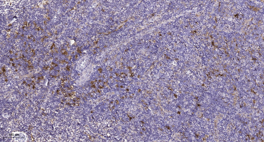
- Immunohistochemical analysis of paraffin-embedded Rat-spleen tissue. 1,PDGFRα Mouse Monoclonal Antibody(7A3) was diluted at 1:200(4°C,overnight). 2, Sodium citrate pH 6.0 was used for antibody retrieval(>98°C,20min). 3,Secondary antibody was diluted at 1:200(room tempeRature, 30min). Negative control was used by secondary antibody only.