Survivin Monoclonal Antibody(Mix)
- Catalog No.:YM3419
- Applications:WB;IHC;IF
- Reactivity:Human;Rat
- Target:
- Survivin
- Fields:
- >>Platinum drug resistance;>>Apoptosis;>>Apoptosis - multiple species;>>Hippo signaling pathway;>>Hepatitis B;>>Pathways in cancer;>>Chemical carcinogenesis - receptor activation;>>Colorectal cancer
- Gene Name:
- BIRC5
- Protein Name:
- Baculoviral IAP repeat-containing protein 5
- Human Gene Id:
- 332
- Human Swiss Prot No:
- O15392
- Mouse Swiss Prot No:
- O70201
- Rat Swiss Prot No:
- Q9JHY7
- Immunogen:
- Recombinant Protein of Survivin
- Specificity:
- The antibody detects endogenous Survivin protein.
- Formulation:
- PBS, pH 7.4, containing 0.5%BSA, 0.02% sodium azide as Preservative and 50% Glycerol.
- Source:
- Monoclonal, Mouse
- Dilution:
- WB 1:1000-2000 IHC 1:200-500. IF 1:50-200
- Purification:
- The antibody was affinity-purified from mouse ascites by affinity-chromatography using epitope-specific immunogen.
- Storage Stability:
- -15°C to -25°C/1 year(Do not lower than -25°C)
- Other Name:
- BIRC5;API4;IAP4;Baculoviral IAP repeat-containing protein 5;Apoptosis inhibitor 4;Apoptosis inhibitor survivin
- Observed Band(KD):
- 16kD
- Background:
- This gene is a member of the inhibitor of apoptosis (IAP) gene family, which encode negative regulatory proteins that prevent apoptotic cell death. IAP family members usually contain multiple baculovirus IAP repeat (BIR) domains, but this gene encodes proteins with only a single BIR domain. The encoded proteins also lack a C-terminus RING finger domain. Gene expression is high during fetal development and in most tumors, yet low in adult tissues. Alternatively spliced transcript variants encoding distinct isoforms have been found for this gene. [provided by RefSeq, Jun 2011],
- Function:
- domain:The BIR repeat is necessary and sufficient for HBXIP binding.,function:May play a role in neoplasia. May counteract a default induction of apoptosis in G2/M phase. Interacts with tubulin. Inhibitor of caspase-3 and caspase-7. Component of the chromosomal passenger complex (CPC), a complex that acts as a key regulator of mitosis. The CPC complex has essential functions at the centromere in ensuring correct chromosome alignment and segregation and is required for chromatin-induced microtubule stabilization and spindle assembly. Isoforms 2 and 3 do not appear to play vital roles in mitosis. Isoform 3 shows a marked reduction in its anti-apoptotic effects when compared with the displayed wild-type isoform.,similarity:Belongs to the IAP family.,similarity:Contains 1 BIR repeat.,subcellular location:Localizes on chromosome arms and inner centromeres from prophase through metaphase and t
- Subcellular Location:
- Cytoplasm . Nucleus . Chromosome . Chromosome, centromere . Cytoplasm, cytoskeleton, spindle . Chromosome, centromere, kinetochore . Midbody . Localizes at the centromeres from prophase to metaphase, at the spindle midzone during anaphase and a the midbody during telophase and cytokinesis. Accumulates in the nucleus upon treatment with leptomycin B (LMB), a XPO1/CRM1 nuclear export inhibitor (By similarity). Localizes on chromosome arms and inner centromeres from prophase through metaphase. Localizes to kinetochores in metaphase, distributes to the midzone microtubules in anaphase and at telophase, localizes exclusively to the midbody (PubMed:11084331). Colocalizes with AURKB at mitotic chromosomes (PubMed:14610074). Acetylation at Lys-129 directs its localization to the nucleus by enhanci
- Expression:
- Expressed only in fetal kidney and liver, and to lesser extent, lung and brain (PubMed:10626797). Abundantly expressed in adenocarcinoma (lung, pancreas, colon, breast, and prostate) and in high-grade lymphomas (PubMed:14741722, PubMed:16329164). Also expressed in various renal cell carcinoma cell lines (PubMed:10626797). Expressed in cochlea including the organ of Corti, the lateral wall, the interdental cells of the Limbus as well as in Schwann cells and cells of the cochlear nerve and the spiral ganglions (at protein level). Not expressed in cells of the inner and outer sulcus or the Reissner's membrane (at protein level) (PubMed:21364656, PubMed:20627126).
- June 19-2018
- WESTERN IMMUNOBLOTTING PROTOCOL
- June 19-2018
- IMMUNOHISTOCHEMISTRY-PARAFFIN PROTOCOL
- June 19-2018
- IMMUNOFLUORESCENCE PROTOCOL
- September 08-2020
- FLOW-CYTOMEYRT-PROTOCOL
- May 20-2022
- Cell-Based ELISA│解您多样本WB检测之困扰
- July 13-2018
- CELL-BASED-ELISA-PROTOCOL-FOR-ACETYL-PROTEIN
- July 13-2018
- CELL-BASED-ELISA-PROTOCOL-FOR-PHOSPHO-PROTEIN
- July 13-2018
- Antibody-FAQs
- Products Images
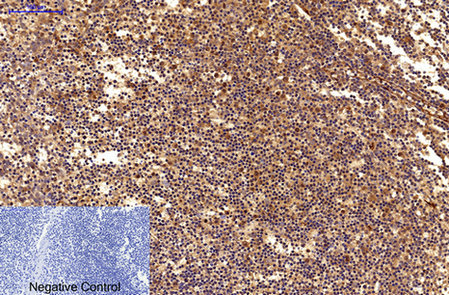
- Immunohistochemical analysis of paraffin-embedded Human-Tonsil tissue. 1,Survivin Monoclonal Antibody(Mix) was diluted at 1:200(4°C,overnight). 2, Sodium citrate pH 6.0 was used for antibody retrieval(>98°C,20min). 3,Secondary antibody was diluted at 1:200(room tempeRature, 30min). Negative control was used by secondary antibody only.

- Immunohistochemical analysis of paraffin-embedded Rat-spinal-cord tissue. 1,Survivin Monoclonal Antibody(Mix) was diluted at 1:200(4°C,overnight). 2, Sodium citrate pH 6.0 was used for antibody retrieval(>98°C,20min). 3,Secondary antibody was diluted at 1:200(room tempeRature, 30min). Negative control was used by secondary antibody only.

- Immunohistochemical analysis of paraffin-embedded Mouse-brain tissue. 1,Survivin Monoclonal Antibody(Mix) was diluted at 1:200(4°C,overnight). 2, Sodium citrate pH 6.0 was used for antibody retrieval(>98°C,20min). 3,Secondary antibody was diluted at 1:200(room tempeRature, 30min). Negative control was used by secondary antibody only.

- Western blot analysis of 1) Hela, 2) 293, 3) PC12 using Survivin Monoclonal Antibody.

- Western blot detection of Survivin in human breast cancer cell line MCF-7(A) and Cal51 (B) using Survivin mouse mAb (YM3419, 1:1000 diluted).Predicted band size: 16kDa.Observed band size:16kDa. Picture was kindly provided by our customer from Tianjin Medical University Cancer Institute and Hospital

- Immunohistochemical analysis of paraffin-embedded Human breast cancer. 1, Using Survivin Mouse mAb (YM3419) diluted at 1:200 (4° overnight). 2, High-pressure and temperature Citric acid, pH6.0 was used for antigen retrieval. 3,Secondary antibody was diluted at 1:200(room temperature, 50min). Picture was kindly provided by our customer from Tianjin Medical University Cancer Institute and Hospital