Total APC Cell-Based Colorimetric ELISA Kit
- Catalog No.:KA3206C
- Applications:ELISA
- Reactivity:Human;Mouse;Rat
- Gene Name:
- APC
- Human Gene Id:
- 324
- Human Swiss Prot No:
- P25054
- Mouse Swiss Prot No:
- Q61315
- Rat Swiss Prot No:
- P70478
- Storage Stability:
- 2-8°C/6 months
- Other Name:
- Adenomatous polyposis coli protein (Protein APC) (Deleted in polyposis 2.5)
- Detection Method:
- Colorimetric
- Background:
- disease:APC mutations have led to some interesting observations. (1) the great majority of the mutations found to date would result in truncation of the APC product. (2) almost all the mutations have occurred within the first half of the coding sequence, and somatic mutations in colorectal tumors are further clustered in a particular region, called MCR (mutation cluster region). (3) most identified point mutations in the APC gene are transitions from cytosine to other nucleotides. (4) the location of germline mutations tends to correlate with the number of colorectal polyps in FAP patients. Inactivation of both alleles of the APC gene seems to be required as an early event to develop most adenomas and carcinomas in the colon and rectum as well as some of those in the stomach.,disease:Defects in APC are a cause of familial adenomatous polyposis (FAP) [MIM:175100]; which includes also Gardner syndrome (GS). FAP and GS contribute to tumor development in patients with uninherited forms of colorectal cancer. FAP is characterized by adenomatous polyps of the colon and rectum, but also of upper gastrointestinal tract (ampullary, duodenal and gastric adenomas). This is a viciously premalignant disease with one or more polyps progressing through dysplasia to malignancy in untreated gene carriers with a median age at diagnosis of 40 years.,disease:Defects in APC are a cause of hereditary desmoid disease (HDD) [MIM:135290]; also called familial infiltrative fibromatosis (FIF). It is an autosomal dominant trait with 100% penetrance and possible variable expression among affected relatives. HDD patients show multifocal fibromatosis of the paraspinal muscles, breast, occiput, arms, lower ribs, abdominal wall, and mesentery. Desmoid tumors appears also as a complication of familial adenomatous polyposis.,disease:Defects in APC are a cause of medulloblastoma (MDB) [MIM:155255]. MDB is a malignant, invasive embryonal tumor of the cerebellum with a preferential manifestation in children. Although the majority of medulloblastomas occur sporadically, some manifest within familial cancer syndromes such as Turcot syndrome and basal cell nevus syndrome (Gorlin syndrome).,disease:Defects in APC are a cause of Turcot syndrome [MIM:276300]. Turcot syndrome is an autosomal dominant disorder characterized by malignant tumors of the brain associated with multiple colorectal adenomas. Skin features include sebaceous cysts, hyperpigmented and cafe au lait spots.,function:Tumor suppressor. Promotes rapid degradation of CTNNB1 and participates in Wnt signaling as a negative regulator. APC activity is correlated with its phosphorylation state.,online information:APC entry,online information:Familial adenomatous polyposis (FAP) website,online information:Information about APC mutations,online information:The Singapore human mutation and polymorphism database,PTM:Phosphorylated by GSK3B.,PTM:Ubiquitinated, leading to its degradation by the proteasome. Ubiquitination is facilitated by Axin. Deubiquitinated by ZRANB1/TRABID.,similarity:Belongs to the adenomatous polyposis coli (APC) family.,similarity:Contains 7 ARM repeats.,subunit:Forms homooligomers. Interacts with DIAPH1 and DIAPH2 (By similarity). Interacts with PDZ domains of DLG1 and DLG3. Associates with catenins. Binds axin. Interacts with the N-terminus of ARHGEF4, and the C-terminus of MAPRE1, MAPRE2 and MAPRE3. Found in a complex consisting of ARHGEF4, APC and CTNNB1. Interacts with APC2.,tissue specificity:Expressed in a variety of tissues.,
- Function:
- cell cycle checkpoint, regulation of cyclin-dependent protein kinase activity, M phase of mitotic cell cycle, mitotic cell cycle, M phase, nuclear division, cytokinesis after mitosis, cell morphogenesis, cell morphogenesis involved in differentiation, cytokinesis, eye development, urogenital system development, cell activation, kidney development,hair follicle development, immune system development, leukocyte differentiation, regulation of myeloid leukocyte differentiation, regionalization, protein complex assembly, negative regulation of protein kinase activity, cell motion,response to DNA damage stimulus, negative regulation of microtubule depolymerization, cell-cell junction assembly,cell cycle, cell cycle arrest, mitosis, regulation of mitosis, mitotic metaphase/anaphase transition, mitotic cell cycle checkpoint, mitotic cell cycle spindle assembly checkpoint, cell adhesion, establis
- Subcellular Location:
- Cell junction, adherens junction . Cytoplasm, cytoskeleton . Cell projection, lamellipodium . Cell projection, ruffle membrane . Cytoplasm . Cell membrane . Associated with the microtubule network at the growing distal tip of microtubules (PubMed:19632184). Accumulates in the lamellipodium and ruffle membrane in response to hepatocyte growth factor (HGF) treatment (PubMed:19151759). The MEMO1-RHOA-DIAPH1 signaling pathway controls localization of the phosphorylated form to the cell membrane (PubMed:20937854). .
- Expression:
- Expressed in a variety of tissues: brain, small intestine, colon, thymus, skeletal muscle, heart, prostate, lung, spleen, ovary, testis kidney, placenta, blood and liver (PubMed:21643010, PubMed:27217144). Isoform 1A: Very strongly expressed in brain but has relatively low expression levels in other tissues (PubMed:19527921, PubMed:21643010, PubMed:27217144). Isoform 1B: Predominant form in all tissues except for brain, including gastric mucosa and blood (PubMed:19527921, PubMed:21643010, PubMed:27217144).
- June 19-2018
- WESTERN IMMUNOBLOTTING PROTOCOL
- June 19-2018
- IMMUNOHISTOCHEMISTRY-PARAFFIN PROTOCOL
- June 19-2018
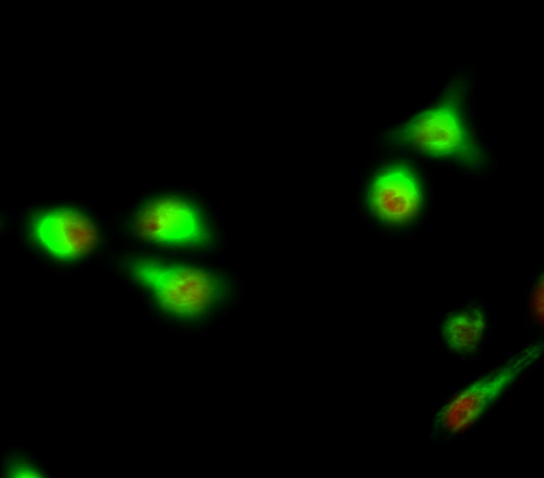
- IMMUNOFLUORESCENCE PROTOCOL
- September 08-2020
- FLOW-CYTOMEYRT-PROTOCOL
- May 20-2022
- Cell-Based ELISA│解您多样本WB检测之困扰
- July 13-2018
- CELL-BASED-ELISA-PROTOCOL-FOR-ACETYL-PROTEIN
- July 13-2018
- CELL-BASED-ELISA-PROTOCOL-FOR-PHOSPHO-PROTEIN
- July 13-2018
- Antibody-FAQs