- 靶点:
- ATG12
- 简介:
- >>FoxO signaling pathway;>>Autophagy - other;>>Autophagy - animal;>>NOD-like receptor signaling pathway;>>RIG-I-like receptor signaling pathway;>>Shigellosis
- 基因名称:
- ATG12 APG12 APG12L
- 蛋白名称:
- ATG12
- Human Gene Id:
- 9140
- Human Swiss Prot No:
- O94817
- Mouse Gene Id:
- 67526
- Mouse Swiss Prot No:
- Q9CQY1
- Rat Gene Id:
- 361321
- Rat Swiss Prot No:
- Q2TBJ5
- 免疫原:
- Synthesized peptide derived from human ATG12 AA range: 80-130
- 特异性:
- This antibody detects endogenous levels of ATG12 at Human/Mouse/Rat
- 组成:
- Liquid in PBS containing 50% glycerol, 0.5% BSA and 0.02% sodium azide.
- 来源:
- Polyclonal, Rabbit,IgG
- 稀释:
- WB 1:500-2000
- 纯化工艺:
- The antibody was affinity-purified from rabbit antiserum by affinity-chromatography using epitope-specific immunogen.
- 浓度:
- 1 mg/ml
- 储存:
- -15°C to -25°C/1 year(Do not lower than -25°C)
- 分子量:
- 15kD
- 背景:
- Autophagy is a process of bulk protein degradation in which cytoplasmic components, including organelles, are enclosed in double-membrane structures called autophagosomes and delivered to lysosomes or vacuoles for degradation. ATG12 is the human homolog of a yeast protein involved in autophagy (Mizushima et al., 1998 [PubMed 9852036]).[supplied by OMIM, Mar 2008],
- 功能:
- function:Required for autophagy.,similarity:Belongs to the ATG12 family.,subunit:Conjugated to ATG5.,tissue specificity:Ubiquitous.,
- 细胞定位:
- Cytoplasm . Preautophagosomal structure membrane ; Peripheral membrane protein . TECPR1 recruits the ATG12-ATG5 conjugate to the autolysosomal membrane.
- 组织表达:
- Ubiquitous.
PTH1-34 promotes osteoblast formation through Beclin1-dependent autophagic activation. JOURNAL OF BONE AND MINERAL METABOLISM J Bone Miner Metab. 2021 Jul;39(4):572-582 WB Mouse MC3T3-E1 cell
货号:YT7272
- June 19-2018
- WESTERN IMMUNOBLOTTING PROTOCOL
- June 19-2018
- IMMUNOHISTOCHEMISTRY-PARAFFIN PROTOCOL
- June 19-2018
- IMMUNOFLUORESCENCE PROTOCOL
- September 08-2020
- FLOW-CYTOMEYRT-PROTOCOL
- May 20-2022
- Cell-Based ELISA│解您多样本WB检测之困扰
- July 13-2018
- CELL-BASED-ELISA-PROTOCOL-FOR-ACETYL-PROTEIN
- July 13-2018
- CELL-BASED-ELISA-PROTOCOL-FOR-PHOSPHO-PROTEIN
- July 13-2018
- Antibody-FAQs
- 产品图片
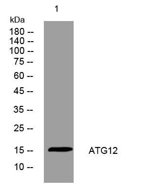
- Western blot analysis of lysates from HpeG2 cells, primary antibody was diluted at 1:1000, 4°over night


