TCP-1 β Monoclonal Antibody
- 货号:YM0613
- 应用:WB;IF;FCM;ELISA
- 种属:Human;Mouse;Rat;Monkey
- 蛋白名称:
- T-complex protein 1 subunit beta
- 免疫原:
- Purified recombinant fragment of human TCP-1 β expressed in E. Coli.
- 特异性:
- TCP-1 β Monoclonal Antibody detects endogenous levels of TCP-1 β protein.
- 组成:
- Liquid in PBS containing 50% glycerol, 0.5% BSA and 0.02% sodium azide.
- 稀释:
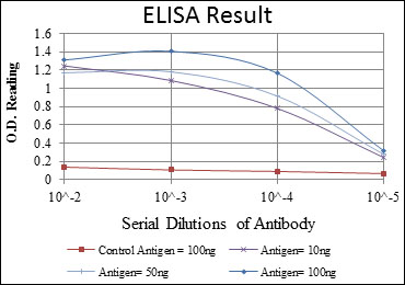
- WB 1:500 - 1:2000. IF 1:200 - 1:1000. Flow cytometry: 1:200 - 1:400. ELISA: 1:10000. Not yet tested in other applications.
- 纯化工艺:
- Affinity purification
- 储存:
- -15°C to -25°C/1 year(Do not lower than -25°C)
- 其他名称:
- CCT2;99D8.1;CCTB;T-complex protein 1 subunit beta;TCP-1-beta;CCT-beta
- 背景:
- The protein encoded by this gene is a molecular chaperone that is a member of the chaperonin containing TCP1 complex (CCT), also known as the TCP1 ring complex (TRiC). This complex consists of two identical stacked rings, each containing eight different proteins. Unfolded polypeptides enter the central cavity of the complex and are folded in an ATP-dependent manner. The complex folds various proteins, including actin and tubulin. Two transcript variants encoding different isoforms have been found for this gene. [provided by RefSeq, Nov 2010],
- 功能:
- function:Molecular chaperone; assist the folding of proteins upon ATP hydrolysis. Known to play a role, in vitro, in the folding of actin and tubulin.,similarity:Belongs to the TCP-1 chaperonin family.,subunit:Heterooligomeric complex of about 850 to 900 kDa that forms two stacked rings, 12 to 16 nm in diameter. Interacts with PACRG.,
- 组织表达:
- B-cell lymphoma,Brain,Cajal-Retzius cell,Fetal brain cortex,Liver,P

- Western Blot analysis using TCP-1 β Monoclonal Antibody against HeLa (1), MCF-7 (2), Jurkat (3), T47D (4), K562 (5), A431 (6), NIH/3T3 (7), PC-12 (8) and Cos7 (9) cell lysate.

- Immunofluorescence analysis of 3T3-L1 cells using TCP-1 β Monoclonal Antibody (green). Blue: DRAQ5 fluorescent DNA dye. Red: Actin filaments have been labeled with Alexa Fluor-555 phalloidin.

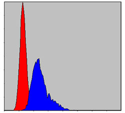
- Flow cytometric analysis of NIH/3T3 cells using TCP-1 β Monoclonal Antibody (blue) and negative control (red).


