PARP-1 Polyclonal Antibody
- Catalog No.:YT6210
- Applications:IHC;IF;WB
- Reactivity:Human;Mouse;Rat
- Target:
- PARP
- Fields:
- >>Base excision repair;>>NF-kappa B signaling pathway;>>Apoptosis;>>Necroptosis;>>Diabetic cardiomyopathy
- Gene Name:
- PARP1 ADPRT PPOL
- Protein Name:
- PARP-1
- Human Gene Id:
- 142
- Human Swiss Prot No:
- P09874
- Immunogen:
- Synthesized peptide derived from human PARP-1. AA range: 410-460
- Specificity:
- This antibody detects endogenous levels of human PARP-1
- Formulation:
- Liquid in PBS containing 50% glycerol, 0.5% BSA and 0.02% sodium azide.
- Source:
- Polyclonal, Rabbit,IgG
- Dilution:
- IHC 1:50-200, WB 1:500-2000. IF 1:50-200
- Purification:
- The antibody was affinity-purified from rabbit antiserum by affinity-chromatography using epitope-specific immunogen.
- Concentration:
- 1 mg/ml
- Storage Stability:
- -15°C to -25°C/1 year(Do not lower than -25°C)
- Other Name:
- Poly [ADP-ribose] polymerase 1 (PARP-1;EC 2.4.2.30;ADP-ribosyltransferase diphtheria toxin-like 1;ARTD1;NAD(+) ADP-ribosyltransferase 1;ADPRT 1;Poly[ADP-ribose] synthase 1)
- Observed Band(KD):
- 113kD
- Background:
- This gene encodes a chromatin-associated enzyme, poly(ADP-ribosyl)transferase, which modifies various nuclear proteins by poly(ADP-ribosyl)ation. The modification is dependent on DNA and is involved in the regulation of various important cellular processes such as differentiation, proliferation, and tumor transformation and also in the regulation of the molecular events involved in the recovery of cell from DNA damage. In addition, this enzyme may be the site of mutation in Fanconi anemia, and may participate in the pathophysiology of type I diabetes. [provided by RefSeq, Jul 2008],
- Function:
- catalytic activity:NAD(+) + (ADP-D-ribosyl)(n)-acceptor = nicotinamide + (ADP-D-ribosyl)(n+1)-acceptor.,function:Involved in the base excision repair (BER) pathway, by catalyzing the poly(ADP-ribosyl)ation of a limited number of acceptor proteins involved in chromatin architecture and in DNA metabolism. This modification follows DNA damages and appears as an obligatory step in a detection/signaling pathway leading to the reparation of DNA strand breaks.,miscellaneous:The ADP-D-ribosyl group of NAD(+) is transferred to an acceptor carboxyl group on a histone or the enzyme itself, and further ADP-ribosyl groups are transferred to the 2'-position of the terminal adenosine moiety, building up a polymer with an average chain length of 20-30 units.,PTM:Phosphorylated by PRKDC. Phosphorylated upon DNA damage, probably by ATM or ATR.,PTM:Poly-ADP-ribosylated by PARP2.,similarity:Contains 1 BRCT
- Subcellular Location:
- Nucleus . Nucleus, nucleolus . Chromosome . Localizes to sites of DNA damage. .
- Expression:
- Brain,Colon carcinoma,Fibroblast,Lung,Ovarian carcinoma,Skin,
The degradation of mixed lineage kinase domain-like protein promotes neuroprotection after ischemic brain injury. Oncotarget Oncotarget. 2017 Sep 15; 8(40): 68393–68401 WB Mouse hemisphere tissue
Synergistic Apoptotic Effect of D-Fraction From Grifola frondosa and Vitamin C on Hepatocellular Carcinoma SMMC-7721 Cells:. INTEGRATIVE CANCER THERAPIES 2016 May 05 WB Human SMMC-7721 cell
Aaptamine attenuates the proliferation and progression of non-small cell lung carcinoma. PHARMACEUTICAL BIOLOGY Pharm Biol. 2020;58(1):1044-1054 WB Human A549 cell,H1299 cell
PARP inhibitor re‑sensitizes Adriamycin resistant leukemia cells through DNA damage and apoptosis. Molecular Medicine Reports Mol Med Rep. 2019 Jan;19(1):75-84 WB Human 1:1000 K562/ADR cell
Marine bromophenol bis (2, 3-dibromo-4, 5-dihydroxybenzyl) ether, represses angiogenesis in HUVEC cells and in zebrafish embryos via inhibiting the VEGF signal systems." Biomedicine & Pharmacotherapy 75 (2015): 58-66.
Coptisine Induces Apoptosis in Human Hepatoma Cells Through Activating 67-kDa Laminin Receptor/cGMP Signaling. Frontiers in Pharmacology 2018 May 18 WB Human 1:500 SMMC7721 cell, HepG2 Cell
High expression of PINK1 promotes proliferation and chemoresistance of NSCLC. ONCOLOGY REPORTS Oncol Rep. 2017 Apr;37(4):2137-2146 WB Human NSCLC tissues A549 cell, H1299 cell
- June 19-2018
- WESTERN IMMUNOBLOTTING PROTOCOL
- June 19-2018
- IMMUNOHISTOCHEMISTRY-PARAFFIN PROTOCOL
- June 19-2018
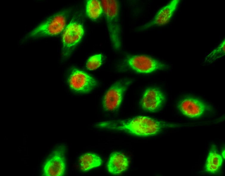
- IMMUNOFLUORESCENCE PROTOCOL
- September 08-2020
- FLOW-CYTOMEYRT-PROTOCOL
- May 20-2022
- Cell-Based ELISA│解您多样本WB检测之困扰
- July 13-2018
- CELL-BASED-ELISA-PROTOCOL-FOR-ACETYL-PROTEIN
- July 13-2018
- CELL-BASED-ELISA-PROTOCOL-FOR-PHOSPHO-PROTEIN
- July 13-2018
- Antibody-FAQs
- Products Images
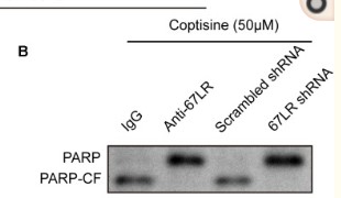
- Zhou, Li, et al. "Coptisine induces apoptosis in human hepatoma cells through activating 67-kDa laminin receptor/cGMP signaling." Frontiers in pharmacology 9 (2018).
- Yao, Chong, et al. "Crocin induces autophagic apoptosis in hepatocellular carcinoma by inhibiting Akt/mTOR activity." OncoTargets and therapy 11 (2018): 2017.
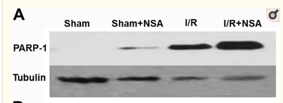
- Zhou, Yanlong, et al. "The degradation of mixed lineage kinase domain-like protein promotes neuroprotection after ischemic brain injury." Oncotarget 8.40 (2017): 68393.
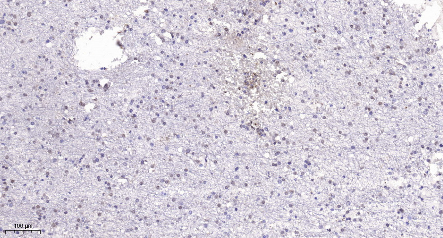
- Immunohistochemical analysis of paraffin-embedded human brain. 1, Tris-EDTA,pH9.0 was used for antigen retrieval. 2 Antibody was diluted at 1:200(4° overnight.3,Secondary antibody was diluted at 1:200(room temperature, 45min).