Tubulin β Polyclonal Antibody
- Catalog No.:YT4780
- Applications:WB;IHC;IF;IP;ELISA
- Reactivity:Human;Mouse;Rat;Pig;Cow;Fish
- Target:
- Tubulin β
- Fields:
- >>Phagosome;>>Gap junction;>>Alzheimer disease;>>Parkinson disease;>>Amyotrophic lateral sclerosis;>>Huntington disease;>>Prion disease;>>Pathways of neurodegeneration - multiple diseases;>>Pathogenic Escherichia coli infection;>>Salmonella infection
- Gene Name:
- TUBB3
- Protein Name:
- Tubulin beta-3 chain
- Human Gene Id:
- 10381
- Human Swiss Prot No:
- Q13509
- Mouse Gene Id:
- 22152
- Mouse Swiss Prot No:
- Q9ERD7
- Rat Gene Id:
- 246118
- Rat Swiss Prot No:
- Q4QRB4
- Immunogen:
- The antiserum was produced against synthesized peptide derived from human Tubulin beta. AA range:401-450
- Specificity:
- Tubulin β Polyclonal Antibody detects endogenous levels of Tubulin β protein.
- Formulation:
- Liquid in PBS containing 50% glycerol, 0.5% BSA and 0.02% sodium azide.
- Source:
- Polyclonal, Rabbit,IgG
- Dilution:
- WB 1:500 - 1:2000. IHC: 1:100-300 ELISA: 1:20000. IF 1:100-300 Not yet tested in other applications.
- Purification:
- The antibody was affinity-purified from rabbit antiserum by affinity-chromatography using epitope-specific immunogen.
- Concentration:
- 1 mg/ml
- Storage Stability:
- -15°C to -25°C/1 year(Do not lower than -25°C)
- Other Name:
- TUBB3;TUBB4;Tubulin beta-3 chain;Tubulin beta-4 chain;Tubulin beta-III
- Observed Band(KD):
- 55kD
- Background:
- tubulin beta 3 class III(TUBB3) Homo sapiens This gene encodes a class III member of the beta tubulin protein family. Beta tubulins are one of two core protein families (alpha and beta tubulins) that heterodimerize and assemble to form microtubules. This protein is primarily expressed in neurons and may be involved in neurogenesis and axon guidance and maintenance. Mutations in this gene are the cause of congenital fibrosis of the extraocular muscles type 3. Alternate splicing results in multiple transcript variants. A pseudogene of this gene is found on chromosome 6. [provided by RefSeq, Oct 2010],
- Function:
- domain:The highly acidic C-terminal region may bind cations such as calcium.,function:Receptor for MSH (alpha, beta and gamma) and ACTH. The activity of this receptor is mediated by G proteins which activate adenylate cyclase.,function:Tubulin is the major constituent of microtubules. It binds two moles of GTP, one at an exchangeable site on the beta chain and one at a non-exchangeable site on the alpha-chain.,polymorphism:Genetic variations in MC1R are associated with variation in skin/hair/eye pigmentation type 2 (SHEP2) [MIM:266300]. Hair, eye and skin pigmentation are among the most visible examples of human phenotypic variation, with a broad normal range that is subject to substantial geographic stratification. In the case of skin, individuals tend to have lighter pigmentation with increasing distance from the equator. By contrast, the majority of variation in human eye and hair col
- Subcellular Location:
- Cytoplasm, cytoskeleton . Cell projection, growth cone . Cell projection, lamellipodium . Cell projection, filopodium .
- Expression:
- Expression is primarily restricted to central and peripheral nervous system. Greatly increased expression in most cancerous tissues.
Molecular characterization and antibacterial immunity functional analysis of the antimicrobial peptide hepcidin from Coregonus ussuriensis berg Fish Shellfish Immun. 2022 Mar;122:78. WB Fish 1:10000 liver
Effects of enriched environment on depression and anxiety-like behavior induced by early life stress: A comparison between different periods. BEHAVIOURAL BRAIN RESEARCH Behav Brain Res. 2021 Aug;411:113389 WB Mouse 1: 800 Hippocampal tissue
Combining network pharmacology, RNA-seq, and metabolomics strategies to reveal the mechanism of Cimicifugae Rhizoma - Smilax glabra Roxb herb pair for the treatment of psoriasis PHYTOMEDICINE Ping Li WB Mouse
Flavonoid-Rich Extract of Oldenlandia diffusa (Willd.) Roxb. Inhibits Gastric Cancer by Activation of Caspase-Dependent Mitochondrial Apoptosis Chinese Journal of Integrative Medicine Qiu-lan Wang WB Human
Suppressing Mesenchymal Stromal Cell Ferroptosis Via Targeting a Metabolism-Epigenetics Axis Corrects their Poor Retention and Insufficient Healing Benefits in the Injured Liver Milieu Advanced Science Fuyang Zhang WB Human,Rat Mesenchymal stromal/stem cells (MSCs),Adipose-derived MSCs (ADSCs)
A reactive oxygen/nitrogen species-eliminating natural product-based nanomedicine for prevention and treatment of inflammatory liver injury. Yu Gao WB Mouse RAW264.7 cell
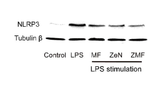
Laxiflorin B covalently binds the tubulin colchicine-binding site to inhibit triple negative breast cancer proliferation and induce apoptosis. CHEMICO-BIOLOGICAL INTERACTIONS Duo Zheng IF,Pull Down Human 1:200 MDA-MB-231 cell,BT549 cell
A network pharmacology integrated serum pharmacochemistry strategy for uncovering efficacy of YXC on hepatocellular carcinoma. JOURNAL OF ETHNOPHARMACOLOGY Hai-bo Cheng WB Human 1:1000 MHCC97H cell,HCCLM3 cell
- June 19-2018
- WESTERN IMMUNOBLOTTING PROTOCOL
- June 19-2018
- IMMUNOHISTOCHEMISTRY-PARAFFIN PROTOCOL
- June 19-2018
- IMMUNOFLUORESCENCE PROTOCOL
- September 08-2020
- FLOW-CYTOMEYRT-PROTOCOL
- May 20-2022
- Cell-Based ELISA│解您多样本WB检测之困扰
- July 13-2018
- CELL-BASED-ELISA-PROTOCOL-FOR-ACETYL-PROTEIN
- July 13-2018
- CELL-BASED-ELISA-PROTOCOL-FOR-PHOSPHO-PROTEIN
- July 13-2018
- Antibody-FAQs
- Products Images
- A reactive oxygen/nitrogen species-eliminating natural product-based nanomedicine for prevention and treatment of inflammatory liver injury. Yu Gao WB Mouse RAW264.7 cell
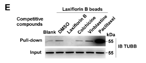
- Laxiflorin B covalently binds the tubulin colchicine-binding site to inhibit triple negative breast cancer proliferation and induce apoptosis. CHEMICO-BIOLOGICAL INTERACTIONS Duo Zheng IF,Pull Down Human 1:200 MDA-MB-231 cell,BT549 cell
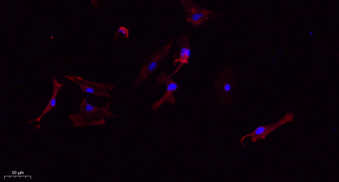
- Immunofluorescence analysis of A549. 1,primary Antibody(red) was diluted at 1:200(4°C overnight). 2, Goat Anti Rabbit IgG (H&L) - Alexa Fluor 594 Secondary antibody was diluted at 1:1000(room temperature, 50min).3, Picture B: DAPI(blue) 10min.
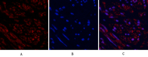
- Immunofluorescence analysis of human-uterus tissue. 1,Tubulin β Polyclonal Antibody(red) was diluted at 1:200(4°C,overnight). 2, Cy3 labled Secondary antibody was diluted at 1:300(room temperature, 50min).3, Picture B: DAPI(blue) 10min. Picture A:Target. Picture B: DAPI. Picture C: merge of A+B
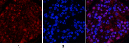
- Immunofluorescence analysis of rat-kidney tissue. 1,Tubulin β Polyclonal Antibody(red) was diluted at 1:200(4°C,overnight). 2, Cy3 labled Secondary antibody was diluted at 1:300(room temperature, 50min).3, Picture B: DAPI(blue) 10min. Picture A:Target. Picture B: DAPI. Picture C: merge of A+B
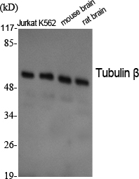
- Western Blot analysis of various cells using Tubulin β Polyclonal Antibody diluted at 1:2000. Secondary antibody(catalog#:RS0002) was diluted at 1:20000
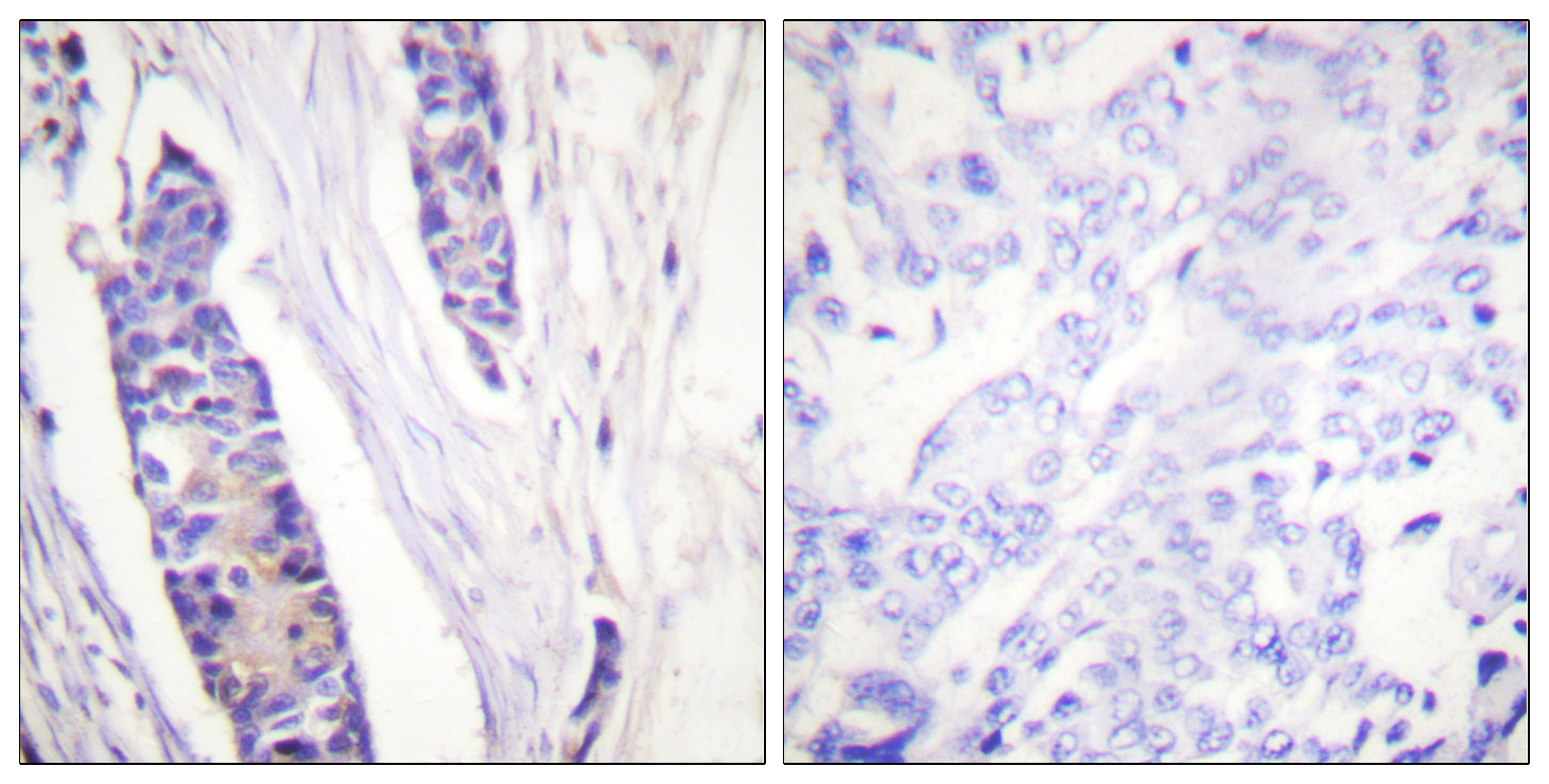
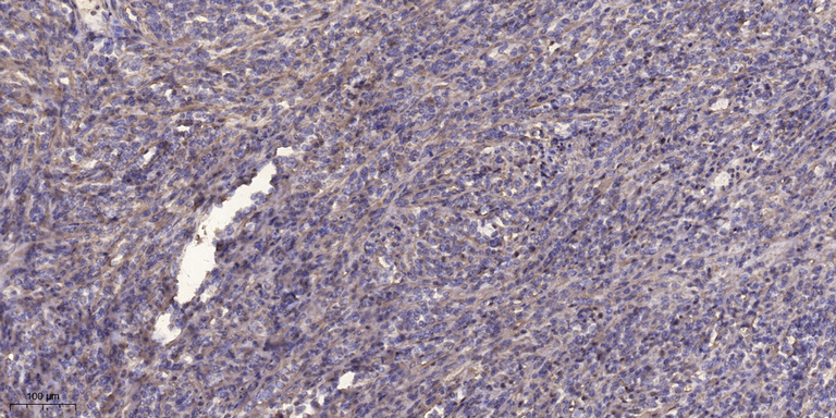
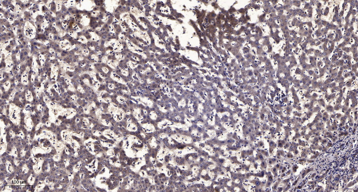
- Immunohistochemistry analysis of paraffin-embedded human breast carcinoma tissue, using Tubulin beta Antibody. The picture on the right is blocked with the synthesized peptide.