TOPBP1 (Phospho Ser1159) rabbit pAb
- Catalog No.:YP1741
- Applications:WB
- Reactivity:Human;Mouse;Rat
- Target:
- TOPBP1
- Fields:
- >>Homologous recombination
- Gene Name:
- TOPBP1 KIAA0259
- Protein Name:
- TOPBP1 (Phospho-Ser1159)
- Human Gene Id:
- 11073
- Human Swiss Prot No:
- Q92547
- Mouse Gene Id:
- 235559
- Mouse Swiss Prot No:
- Q6ZQF0
- Immunogen:
- Synthesized peptide derived from human TOPBP1 (Phospho-Ser1159)
- Specificity:
- This antibody detects endogenous levels of TOPBP1 (Phospho-Ser1159) at Human, Mouse,Rat
- Formulation:
- Liquid in PBS containing 50% glycerol, 0.5% BSA and 0.02% sodium azide.
- Source:
- Polyclonal, Rabbit,IgG
- Dilution:
- WB 1:500-2000
- Purification:
- The antibody was affinity-purified from rabbit serum by affinity-chromatography using specific immunogen.
- Concentration:
- 1 mg/ml
- Storage Stability:
- -15°C to -25°C/1 year(Do not lower than -25°C)
- Other Name:
- DNA topoisomerase 2-binding protein 1 (DNA topoisomerase II-beta-binding protein 1) (TopBP1) (DNA topoisomerase II-binding protein 1)
- Molecular Weight(Da):
- 167kD
- Background:
- This gene encodes a binding protein which interacts with the C-terminal region of topoisomerase II beta. This interaction suggests a supportive role for this protein in the catalytic reactions of topoisomerase II beta through transient breakages of DNA strands. [provided by RefSeq, Jul 2008],
- Function:
- function:Required for DNA replication. Plays a role in the rescue of stalled replication forks and checkpoint control. Binds double-stranded DNA breaks and nicks as well as single-stranded DNA. Recruits the SWI/SNF chromatin remodeling complex to E2F1-responsive promoters. Down-regulates E2F1 activity and inhibits E2F1-dependent apoptosis during G1/S transition and after DNA damage. Induces a large increase in the kinase activity of ATR.,induction:Up-regulated during the S phase of the cell cycle. Up-regulated by E2F1 and interferon.,PTM:Phosphorylated on serine and threonine residues in response to X-ray irradiation.,PTM:Ubiquitinated and degraded by the proteasome. X-ray irradiation reduces ubiquitination.,similarity:Contains 8 BRCT domains.,subcellular location:Detected on unpaired autosomes in meiotic prophase cells. Detected on X and Y chromosomes during later stages of prophase. Co
- Subcellular Location:
- Nucleus. Cytoplasm, cytoskeleton, microtubule organizing center, centrosome. Cytoplasm, cytoskeleton, spindle pole. Chromosome. Detected on unpaired autosomes in meiotic prophase cells. Detected on X and Y chromosomes during later stages of prophase. Colocalizes with ATR and H2AX at unsynapsed chromosome cores during prophase (By similarity). Has a uniform nuclear distribution during G phase. Colocalizes with BRCA1 at stalled replication forks during S phase. In mitotic cells it colocalizes with BRCA1 at spindle poles and centrosomes during metaphase and anaphase. Detected in discrete foci together with PML and numerous DNA repair enzymes after DNA damage by alkylating agents, UV or gamma irradiation. Localizes to sites of DNA damage in a H2AX- independent manner. .
- Expression:
- Highly expressed in heart, brain, placenta, lung and kidney.
- June 19-2018
- WESTERN IMMUNOBLOTTING PROTOCOL
- June 19-2018
- IMMUNOHISTOCHEMISTRY-PARAFFIN PROTOCOL
- June 19-2018
- IMMUNOFLUORESCENCE PROTOCOL
- September 08-2020
- FLOW-CYTOMEYRT-PROTOCOL
- May 20-2022
- Cell-Based ELISA│解您多样本WB检测之困扰
- July 13-2018
- CELL-BASED-ELISA-PROTOCOL-FOR-ACETYL-PROTEIN
- July 13-2018
- CELL-BASED-ELISA-PROTOCOL-FOR-PHOSPHO-PROTEIN
- July 13-2018
- Antibody-FAQs
- Products Images

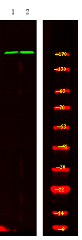
- Western Blot analysis of 1HeLa cell 2 LPS 100ng/mL 30min treated ,using primary antibody at 1:1000 dilution. Secondary antibody(catalog#:RS23920) was diluted at 1:10000

