Stat1 (phospho Tyr701) Polyclonal Antibody
- Catalog No.:YP0249
- Applications:IF;WB;IHC;IP;ELISA
- Reactivity:Human;Mouse;Rat;Monkey
- Target:
- Stat1
- Fields:
- >>Chemokine signaling pathway;>>Necroptosis;>>Osteoclast differentiation;>>Toll-like receptor signaling pathway;>>NOD-like receptor signaling pathway;>>C-type lectin receptor signaling pathway;>>JAK-STAT signaling pathway;>>Th1 and Th2 cell differentiation;>>Th17 cell differentiation;>>Prolactin signaling pathway;>>Thyroid hormone signaling pathway;>>AGE-RAGE signaling pathway in diabetic complications;>>Growth hormone synthesis, secretion and action;>>Leishmaniasis;>>Toxoplasmosis;>>Tuberculosis;>>Hepatitis C;>>Hepatitis B;>>Measles;>>Influenza A;>>Human papillomavirus infection;>>Kaposi sarcoma-associated herpesvirus infection;>>Herpes simplex virus 1 infection;>>Epstein-Barr virus infection;>>Coronavirus disease - COVID-19;>>Pathways in cancer;>>Pancreatic cancer;>>PD-L1 expression and PD-1 checkpoint pathway in cancer;>>Inflammatory bowel disease
- Gene Name:
- STAT1
- Protein Name:
- Signal transducer and activator of transcription 1-alpha/beta
- Human Gene Id:
- 6772
- Human Swiss Prot No:
- P42224
- Mouse Swiss Prot No:
- P42225
- Immunogen:
- The antiserum was produced against synthesized peptide derived from human STAT1 around the phosphorylation site of Tyr701. AA range:668-717
- Specificity:
- Phospho-Stat1 (Y701) Polyclonal Antibody detects endogenous levels of Stat1 protein only when phosphorylated at Y701.
- Formulation:
- Liquid in PBS containing 50% glycerol, 0.5% BSA and 0.02% sodium azide.
- Source:
- Polyclonal, Rabbit,IgG
- Dilution:
- IF 1:50-200 WB 1:500 - 1:2000. IHC 1:100 - 1:300. Immunoprecipitation: 2-5 ug:mg lysate. ELISA: 1:10000. Not yet tested in other applications.
- Purification:
- The antibody was affinity-purified from rabbit antiserum by affinity-chromatography using epitope-specific immunogen.
- Concentration:
- 1 mg/ml
- Storage Stability:
- -15°C to -25°C/1 year(Do not lower than -25°C)
- Other Name:
- STAT1;Signal transducer and activator of transcription 1-alpha/beta;Transcription factor ISGF-3 components p91/p84
- Observed Band(KD):
- 87kD
- Background:
- The protein encoded by this gene is a member of the STAT protein family. In response to cytokines and growth factors, STAT family members are phosphorylated by the receptor associated kinases, and then form homo- or heterodimers that translocate to the cell nucleus where they act as transcription activators. This protein can be activated by various ligands including interferon-alpha, interferon-gamma, EGF, PDGF and IL6. This protein mediates the expression of a variety of genes, which is thought to be important for cell viability in response to different cell stimuli and pathogens. Two alternatively spliced transcript variants encoding distinct isoforms have been described. [provided by RefSeq, Jul 2008],
- Function:
- disease:Defects in STAT1 are a cause of mendelian susceptibility to mycobacterial disease (MSMD) [MIM:209950]; also known as familial disseminated atypical mycobacterial infection. This rare condition confers predisposition to illness caused by moderately virulent mycobacterial species, such as Bacillus Calmette-Guerin (BCG) vaccine and environmental non-tuberculous mycobacteria, and by the more virulent Mycobacterium tuberculosis. Other microorganisms rarely cause severe clinical disease in individuals with susceptibility to mycobacterial infections, with the exception of Salmonella which infects less than 50% of these individuals. The pathogenic mechanism underlying MSMD is the impairment of interferon-gamma mediated immunity whose severity determines the clinical outcome. Some patients die of overwhelming mycobacterial disease with lepromatous-like lesions in early childhood, whereas
- Subcellular Location:
- Cytoplasm . Nucleus . Translocated into the nucleus upon tyrosine phosphorylation and dimerization, in response to IFN-gamma and signaling by activated FGFR1, FGFR2, FGFR3 or FGFR4 (PubMed:15322115). Monomethylation at Lys-525 is required for phosphorylation at Tyr-701 and translocation into the nucleus (PubMed:28753426). Translocates into the nucleus in response to interferon-beta stimulation (PubMed:26479788). .
- Expression:
- B-cell,Brain,Retina,Testis,
Suppression of the hyaluronic acid pathway induces M1 macrophages polarization via STAT1 in glioblastoma. Cell Death Discovery2022 Apr;8(1):1-13. Human U937 monocytes,U251 cell,LN229 cell
Integrated Network Pharmacology and Metabolomics Analysis to Reveal the Potential Mechanism of Siwu Paste on Aplastic Anemia Induced by Chemotherapy Drugs. Drug Design Development and Therapy2022; 16: 1231–1254. Rat BMCs
Polysaccharides From the Aerial Parts of Tetrastigma Hemsleyanum Diels et Gilg Induce Bidirectional Immunity and Ameliorate LPS-Induced Acute Respiratory Distress Syndrome in Mice. Front Pharmacol. 2022 Mar;13:838873-838873. WB Mouse 1:1000 lung
Quantitative Proteomic Analysis Reveals That Arctigenin Alleviates Concanavalin A-Induced Hepatitis Through Suppressing Immune System and Regulating Autophagy. Frontiers in Immunology 2018 Aug 16 WB Mouse liver
gga-miR-27b-3p enhances type I interferon expression and suppresses infectious bursal disease virus replication via targeting cellular suppressors of cytokine signaling 3 and 6 (SOCS3 and 6). VIRUS RESEARCH Virus Res. 2020 May;281:197910 WB Chicken DF-1 cell
Quantitative Proteomic Analysis Reveals That Arctigenin Alleviates Concanavalin A-Induced Hepatitis Through Suppressing Immune System and Regulating Autophagy. Frontiers in Immunology 2018 Aug 16 WB Mouse liver
Effect of TRPM8 Functional Loss on Corneal Epithelial Wound Healing in Mice INVESTIGATIVE OPHTHALMOLOGY & VISUAL SCIENCE Lixin Xie IF Mouse Corneal
- June 19-2018
- WESTERN IMMUNOBLOTTING PROTOCOL
- June 19-2018
- IMMUNOHISTOCHEMISTRY-PARAFFIN PROTOCOL
- June 19-2018
- IMMUNOFLUORESCENCE PROTOCOL
- September 08-2020
- FLOW-CYTOMEYRT-PROTOCOL
- May 20-2022
- Cell-Based ELISA│解您多样本WB检测之困扰
- July 13-2018
- CELL-BASED-ELISA-PROTOCOL-FOR-ACETYL-PROTEIN
- July 13-2018
- CELL-BASED-ELISA-PROTOCOL-FOR-PHOSPHO-PROTEIN
- July 13-2018
- Antibody-FAQs
- Products Images

- Immunofluorescence analysis of Hela cell. 1,Stat1 (phospho Tyr701) Polyclonal Antibody(red) was diluted at 1:200(4° overnight). HER2 Monoclonal Antibody(11H9)(green) was diluted at 1:200(4° overnight). 2, Goat Anti Rabbit Alexa Fluor 594 Catalog:RS3611 was diluted at 1:1000(room temperature, 50min). Goat Anti Mouse Alexa Fluor 488 Catalog:RS3208 was diluted at 1:1000(room temperature, 50min).
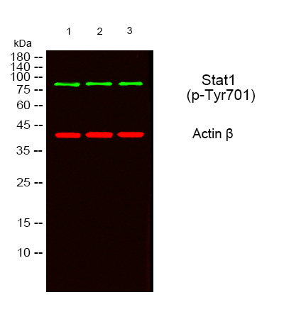
- Western blot analysis of lysates from 1) MOUSE-BRAIN, 2) SY-SY5X ,3) COS7 cells, (Green) primary antibody was diluted at 1:1000, 4°over night, secondary antibody(cat:RS23920)was diluted at 1:10000, 37° 1hour. (Red) Actin β Monoclonal Antibody(5B7) (cat:YM3028) antibody was diluted at 1:5000 as loading control, 4° over night,secondary antibody(cat:RS23710)was diluted at 1:10000, 37° 1hour.
-if-100.jpg)
- Immunofluorescence analysis of rat-spleen tissue. 1,Stat1 (phospho Tyr701) Polyclonal Antibody(red) was diluted at 1:200(4°C,overnight). 2, Cy3 labled Secondary antibody was diluted at 1:300(room temperature, 50min).3, Picture B: DAPI(blue) 10min. Picture A:Target. Picture B: DAPI. Picture C: merge of A+B
-if-101.jpg)
- Immunofluorescence analysis of rat-spleen tissue. 1,Stat1 (phospho Tyr701) Polyclonal Antibody(red) was diluted at 1:200(4°C,overnight). 2, Cy3 labled Secondary antibody was diluted at 1:300(room temperature, 50min).3, Picture B: DAPI(blue) 10min. Picture A:Target. Picture B: DAPI. Picture C: merge of A+B
-if-102.jpg)
- Immunofluorescence analysis of mouse-spleen tissue. 1,Stat1 (phospho Tyr701) Polyclonal Antibody(red) was diluted at 1:200(4°C,overnight). 2, Cy3 labled Secondary antibody was diluted at 1:300(room temperature, 50min).3, Picture B: DAPI(blue) 10min. Picture A:Target. Picture B: DAPI. Picture C: merge of A+B
poly-ihc-mouse-liver.jpg)
- Immunohistochemical analysis of paraffin-embedded Mouse-liver tissue. 1,Stat1 (phospho Tyr701) Polyclonal Antibody was diluted at 1:200(4°C,overnight). 2, Sodium citrate pH 6.0 was used for antibody retrieval(>98°C,20min). 3,Secondary antibody was diluted at 1:200(room tempeRature, 30min). Negative control was used by secondary antibody only.
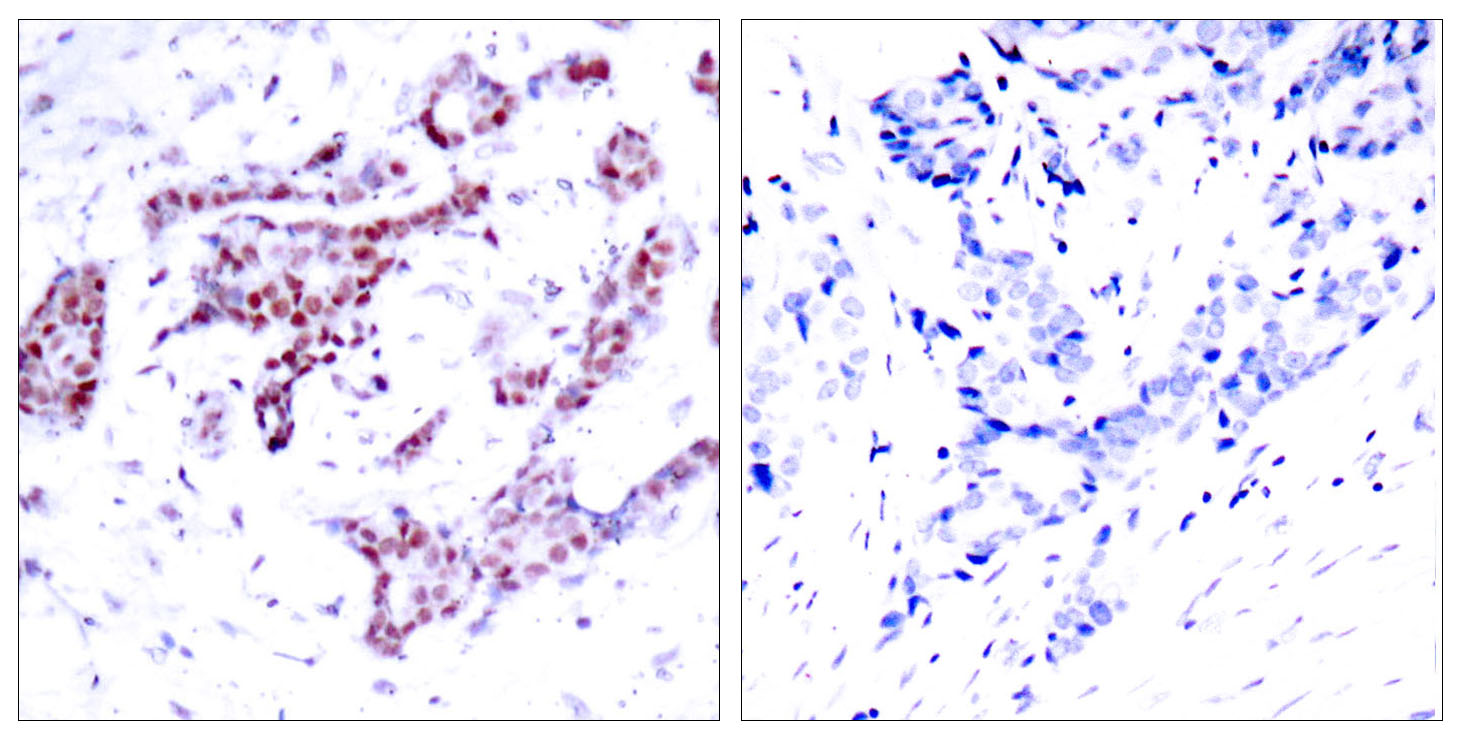
- Immunohistochemistry analysis of paraffin-embedded human breast carcinoma, using STAT1 (Phospho-Tyr701) Antibody. The picture on the right is blocked with the phospho peptide.
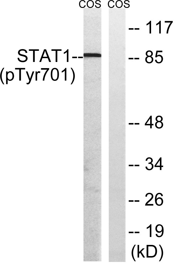
- Western blot analysis of lysates from COS7 cells, using STAT1 (Phospho-Tyr701) Antibody. The lane on the right is blocked with the phospho peptide.
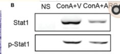
- Feng, Qin. "Quantitative proteomic analysis reveals that Arctigenin alleviates concanavalin A-induced hepatitis through suppressing immune system and regulating autophagy." Frontiers in immunology 9 (2018): 1881.

