Aurora A (PT0150R) PT® Rabbit mAb
- Catalog No.:YM8087
- Applications:WB;IHC;IF;IP;ELISA
- Reactivity:Human;
- Target:
- Aurora A
- Fields:
- >>Oocyte meiosis;>>Progesterone-mediated oocyte maturation
- Gene Name:
- AURKA AIK AIRK1 ARK1 AURA AYK1 BTAK IAK1 STK15 STK6
- Protein Name:
- Aurora A
- Human Gene Id:
- 6790
- Human Swiss Prot No:
- O14965
- Mouse Gene Id:
- 20878
- Mouse Swiss Prot No:
- P97477
- Rat Swiss Prot No:
- P59241
- Specificity:
- endogenous
- Formulation:
- PBS, 50% glycerol, 0.05% Proclin 300, 0.05%BSA
- Source:
- Monoclonal, rabbit, IgG, Kappa
- Dilution:
- IHC 1:200-1000,WB 1:1000-5000,IF 1:200-1000,ELISA 1:5000-20000,IP 1:50-200
- Purification:
- Protein A
- Storage Stability:
- -15°C to -25°C/1 year(Do not lower than -25°C)
- Other Name:
- Aurora kinase A (EC 2.7.11.1) (Aurora 2) (Aurora/IPL1-related kinase 1) (ARK-1) (Aurora-related kinase 1) (hARK1) (Breast tumor-amplified kinase) (Serine/threonine-protein kinase 15) (Serine/threonine-protein kinase 6) (Serine/threonine-protein kinase aurora-A)
- Molecular Weight(Da):
- 45kD
- Observed Band(KD):
- 45kD
- Background:
- Aurora A(AURKA) Homo sapiens The protein encoded by this gene is a cell cycle-regulated kinase that appears to be involved in microtubule formation and/or stabilization at the spindle pole during chromosome segregation. The encoded protein is found at the centrosome in interphase cells and at the spindle poles in mitosis. This gene may play a role in tumor development and progression. A processed pseudogene of this gene has been found on chromosome 1, and an unprocessed pseudogene has been found on chromosome 10. Multiple transcript variants encoding the same protein have been found for this gene.
- Subcellular Location:
- Membranous
- Expression:
- Highly expressed in testis and weakly in skeletal muscle, thymus and spleen. Also highly expressed in colon, ovarian, prostate, neuroblastoma, breast and cervical cancer cell lines.
- June 19-2018
- WESTERN IMMUNOBLOTTING PROTOCOL
- June 19-2018
- IMMUNOHISTOCHEMISTRY-PARAFFIN PROTOCOL
- June 19-2018
- IMMUNOFLUORESCENCE PROTOCOL
- September 08-2020
- FLOW-CYTOMEYRT-PROTOCOL
- May 20-2022
- Cell-Based ELISA│解您多样本WB检测之困扰
- July 13-2018
- CELL-BASED-ELISA-PROTOCOL-FOR-ACETYL-PROTEIN
- July 13-2018
- CELL-BASED-ELISA-PROTOCOL-FOR-PHOSPHO-PROTEIN
- July 13-2018
- Antibody-FAQs
- Products Images

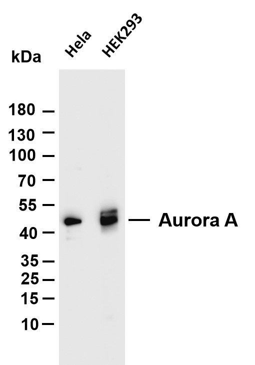
- Various whole cell lysates were separated by 4-20% SDS-PAGE, and the membrane was blotted with anti-Aurora A (PT0150R) antibody. The HRP-conjugated Goat anti-Rabbit IgG(H + L) antibody was used to detect the antibody. Lane 1: Hela Lane 2: Mouse brain Lane 3: Rat brain Predicted band size: 45kDa Observed band size: 45kDa

