SMC1 Monoclonal Antibody
- Catalog No.:YM1095
- Applications:WB;IHC;IF;FCM
- Reactivity:Human;Mouse;Rat;Pig
- Target:
- SMC1
- Fields:
- >>Cell cycle;>>Oocyte meiosis
- Gene Name:
- SMC1A
- Protein Name:
- Structural maintenance of chromosomes protein 1A
- Human Gene Id:
- 8243
- Human Swiss Prot No:
- Q14683
- Mouse Gene Id:
- 24061
- Mouse Swiss Prot No:
- Q9CU62
- Rat Gene Id:
- 63996
- Rat Swiss Prot No:
- Q9Z1M9
- Immunogen:
- Purified recombinant human SMC1 (N-terminus) protein fragments expressed in E.coli.
- Specificity:
- SMC1 Monoclonal Antibody detects endogenous levels of SMC1 protein.
- Formulation:
- Liquid in PBS containing 50% glycerol, 0.5% BSA and 0.02% sodium azide.
- Source:
- Monoclonal, Mouse
- Dilution:
- WB 1:1000 - 1:2000. IHC 1:500 - 1:1000. Flow cytometry: 1:100 - 1:200.. IF 1:50-200
- Purification:
- Affinity purification
- Concentration:
- 1 mg/ml
- Storage Stability:
- -15°C to -25°C/1 year(Do not lower than -25°C)
- Other Name:
- SMC1A;DXS423E;KIAA0178;SB1.8;SMC1;SMC1L1;Structural maintenance of chromosomes protein 1A;SMC protein 1A;SMC-1-alpha;SMC-1A;Sb1.8
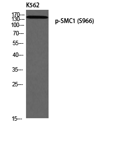
- Molecular Weight(Da):
- 143kD
- Background:
- structural maintenance of chromosomes 1A(SMC1A) Homo sapiens Proper cohesion of sister chromatids is a prerequisite for the correct segregation of chromosomes during cell division. The cohesin multiprotein complex is required for sister chromatid cohesion. This complex is composed partly of two structural maintenance of chromosomes (SMC) proteins, SMC3 and either SMC1B or the protein encoded by this gene. Most of the cohesin complexes dissociate from the chromosomes before mitosis, although those complexes at the kinetochore remain. Therefore, the encoded protein is thought to be an important part of functional kinetochores. In addition, this protein interacts with BRCA1 and is phosphorylated by ATM, indicating a potential role for this protein in DNA repair. This gene, which belongs to the SMC gene family, is located in an area of the X-chromosome that escapes X inactivation. Mutations in this gene result in Cornelia de Lange syndrome. Altern
- Function:
- disease:Defects in SMC1A are the cause of Cornelia de Lange syndrome type 2 (CDLS2) [MIM:300590]; also known as Cornelia de Lange syndrome X-linked. CDLS is a clinically heterogeneous developmental disorder associated with malformations affecting multiple systems. CDLS is characterized by facial dysmorphisms, abnormal hands and feet, growth delay, cognitive retardation and various other malformations including gastroesophageal dysfunction and cardiac, ophthalmologic and genitourinary anomalies.,domain:The flexible hinge domain, which separates the large intramolecular coiled coil regions, allows the heterotypic interaction with the corresponding domain of SMC3, forming a V-shaped heterodimer. The two heads of the heterodimer are then connected by different ends of the cleavable RAD21 protein, forming a ring structure.,function:Involved in chromosome cohesion during cell cycle and in DNA
- Subcellular Location:
- Nucleus . Chromosome . Chromosome, centromere, kinetochore . Associates with chromatin. Before prophase it is scattered along chromosome arms. During prophase, most of cohesin complexes dissociate from chromatin probably because of phosphorylation by PLK, except at centromeres, where cohesin complexes remain. At anaphase, the RAD21 subunit of the cohesin complex is cleaved, leading to the dissociation of the complex from chromosomes, allowing chromosome separation. In germ cells, cohesin complex dissociates from chromatin at prophase I, and may be replaced by a meiosis-specific cohesin complex. The phosphorylated form on Ser-957 and Ser-966 associates with chromatin during G1/S/G2 phases but not during M phase, suggesting that phosphorylation does not regulate cohesin function. Integral co
- Expression:
- Aorta,Bone marrow,Brain,Epithelium,Fibroblast,Testis,Uterus endothe
- June 19-2018
- WESTERN IMMUNOBLOTTING PROTOCOL
- June 19-2018
- IMMUNOHISTOCHEMISTRY-PARAFFIN PROTOCOL
- June 19-2018
- IMMUNOFLUORESCENCE PROTOCOL
- September 08-2020
- FLOW-CYTOMEYRT-PROTOCOL
- May 20-2022
- Cell-Based ELISA│解您多样本WB检测之困扰
- July 13-2018
- CELL-BASED-ELISA-PROTOCOL-FOR-ACETYL-PROTEIN
- July 13-2018
- CELL-BASED-ELISA-PROTOCOL-FOR-PHOSPHO-PROTEIN
- July 13-2018
- Antibody-FAQs
- Products Images
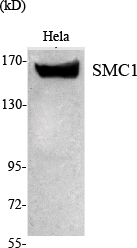
- Western Blot analysis using SMC1 Monoclonal Antibody against HeLa nuclear extract.
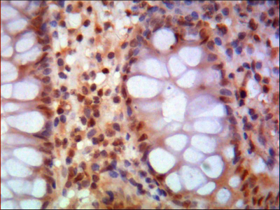
- Immunohistochemistry analysis of paraffin-embedded human colon using SMC1 Monoclonal Antibody.

- Flow cytometric analysis of HeLa cells stained with SMC1 Monoclonal Antibody (red), followed by FITC-conjugated goat anti-mouse IgG. Black line histogram represents the isotype control, normal mouse IgG.