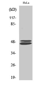
Total VASP Cell-Based Colorimetric ELISA Kit
- Catalog No.:KA4315C
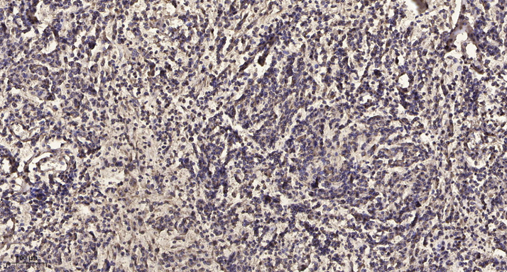
- Applications:ELISA
- Reactivity:Human;Mouse;Rat
- Gene Name:
- VASP
- Human Gene Id:
- 7408
- Human Swiss Prot No:
- P50552
- Mouse Swiss Prot No:
- P70460
- Storage Stability:
- 2-8°C/6 months
- Other Name:
- Vasodilator-stimulated phosphoprotein (VASP)
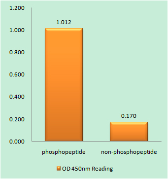
- Detection Method:
- Colorimetric
- Background:
- domain:The EVH2 domain is comprised of 3 regions. Block A is a thymosin-like domain required for G-actin binding. The KLKR motif within this block is essential for the G-actin binding and for actin polymerization. Block B is required for F-actin binding and subcellular location, and Block C for tetramerization.,domain:The WH1 domain mediates interaction with XIRP1.,function:Ena/VASP proteins are actin-associated proteins involved in a range of processes dependent on cytoskeleton remodeling and cell polarity such as axon guidance and lamellipodial and filopodial dynamics in migrating cells. VASP promotes actin nucleation and increases the rate of actin polymerization in the presence of capping protein. Plays a role in actin-based activity of Listeria monocytogenes in platelets.,PTM:Major substrate for cAMP-dependent (PKA) and cGMP-dependent protein kinase (PKG) in platelets. The preferred site for PKA is Ser-157, the preferred site for PKG, Ser-239. In ADP-activated platelets, phosphorylation by PKA or PKG on Ser-157 leads to fibrinogen receptor inhibition. Phosphorylation on Thr-278 requires prior phosphorylation on Ser-157 and Ser-239. In response to phorbol ester (PMA) stimulation, phosphorylated by PKC/PRKCA. In response to thrombin, phosphorylated by both PKC and ROCK1.,similarity:Belongs to the Ena/VASP family.,similarity:Contains 1 WH1 domain.,subcellular location:Targeted to stress fibers and focal adhesions through interaction with a number of proteins including MRL family members. Localizes to the plasma membrane in protruding lamellipodia and filopodial tips. Stimulation by thrombin or PMA, also translocates VASP to focal adhesions.,subunit:Homotetramer. Interacts with PFN1, PFN2, LPP, ACTN1 and ACTG1. Interacts, via the EVH1, with the Pro-rich regions of ZYX. This interaction is important for targeting to focal adhesions and the formation of actin-rich structures at the apical surface of cells. Interacts, via the EVH1 domain, with the Pro-rich domain of Listeria monocytogenes actA. Interacts with APBB1IP. Interacts, via the Pro-rich domain, with the C-terminal SH3 domain of DNMBP.,tissue specificity:Highly expressed in platelets.,
- Function:
- cell morphogenesis, cell morphogenesis involved in differentiation, embryonic epithelial tube formation, neural tube formation, neural tube closure, morphogenesis of an epithelium, cell motion, cytoskeleton organization, axonogenesis,axon guidance, embryonic development ending in birth or egg hatching, primary neural tube formation,morphogenesis of embryonic epithelium, neural tube development, actin filament-based process, cell projection organization, actin cytoskeleton organization, neuron differentiation, neuron projection development, cellular component morphogenesis, cell part morphogenesis, tube lumen formation, tube morphogenesis, tube development,chordate embryonic development, embryonic morphogenesis, neuron development, cell morphogenesis involved in neuron differentiation, tissue morphogenesis, neuron projection morphogenesis, cell projection morphogenesis,epithelium developm
- Subcellular Location:
- Cytoplasm. Cytoplasm, cytoskeleton. Cell junction, focal adhesion. Cell junction, tight junction . Cell projection, lamellipodium membrane. Cell projection, filopodium membrane. Targeted to stress fibers and focal adhesions through interaction with a number of proteins including MRL family members. Localizes to the plasma membrane in protruding lamellipodia and filopodial tips. Stimulation by thrombin or PMA, also translocates VASP to focal adhesions. Localized along the sides of actin filaments throughout the peripheral cytoplasm under basal conditions. In pre-apoptotic cells, colocalizes with MEFV in large specks (pyroptosomes).
- Expression:
- Highly expressed in platelets.
- June 19-2018
- WESTERN IMMUNOBLOTTING PROTOCOL
- June 19-2018
- IMMUNOHISTOCHEMISTRY-PARAFFIN PROTOCOL
- June 19-2018
- IMMUNOFLUORESCENCE PROTOCOL
- September 08-2020
- FLOW-CYTOMEYRT-PROTOCOL
- May 20-2022
- Cell-Based ELISA│解您多样本WB检测之困扰
- July 13-2018
- CELL-BASED-ELISA-PROTOCOL-FOR-ACETYL-PROTEIN
- July 13-2018
- CELL-BASED-ELISA-PROTOCOL-FOR-PHOSPHO-PROTEIN
- July 13-2018
- Antibody-FAQs