Topoisomerase IIα (ABT272) IHC kit
- Catalog No.:IHCM6914
- Applications:IHC
- Reactivity:Human;Mouse;Rat;
- Target:
- Topo IIα
- Fields:
- >>Platinum drug resistance
- Gene Name:
- TOP2A TOP2
- Protein Name:
- Topoisomerase IIα
- Human Gene Id:
- 7153
- Human Swiss Prot No:
- P11388
- Immunogen:
- Synthesized peptide derived from human Topoisomerase IIα AA range: 1400-1531
- Specificity:
- The antibody can specifically recognize human Topoisomerase IIα protein.
- Source:
- Mouse, Monoclonal/IgG1, kappa
- Purification:
- The antibody was affinity-purified from ascites by affinity-chromatography using specific immunogen.
- Storage Stability:
- 2°C to 8°C/1 year
- Other Name:
- DNA topoisomerase 2-alpha (EC 5.99.1.3;DNA topoisomerase II, alpha isozyme)
- Background:
- This gene encodes a DNA topoisomerase, an enzyme that controls and alters the topologic states of DNA during transcription. This nuclear enzyme is involved in processes such as chromosome condensation, chromatid separation, and the relief of torsional stress that occurs during DNA transcription and replication. It catalyzes the transient breaking and rejoining of two strands of duplex DNA which allows the strands to pass through one another, thus altering the topology of DNA. Two forms of this enzyme exist as likely products of a gene duplication event. The gene encoding this form, alpha, is localized to chromosome 17 and the beta gene is localized to chromosome 3. The gene encoding this enzyme functions as the target for several anticancer agents and a variety of mutations in this gene have been associated with the development of drug resistance. Reduced activity of this enzyme may also pla
- Function:
- catalytic activity:ATP-dependent breakage, passage and rejoining of double-stranded DNA.,enzyme regulation:Specifically inhibited by the intercalating agent amsacrine.,function:Control of topological states of DNA by transient breakage and subsequent rejoining of DNA strands. Topoisomerase II makes double-strand breaks.,miscellaneous:Eukaryotic topoisomerase I and II can relax both negative and positive supercoils, whereas prokaryotic enzymes relax only negative supercoils.,PTM:Phosphorylation has no effect on catalytic activity.,similarity:Belongs to the type II topoisomerase family.,subcellular location:Generally located in the nucleoplasm.,subunit:Homodimer. Interacts with COPS5.,
- Subcellular Location:
- Nuclear
- Expression:
- Expressed in the tonsil, spleen, lymph node, thymus, skin, pancreas, testis, colon, kidney, liver, brain and lung (PubMed:9155056). Also found in high-grade lymphomas, squamous cell lung tumors and seminomas (PubMed:9155056).
- June 19-2018
- WESTERN IMMUNOBLOTTING PROTOCOL
- June 19-2018
- IMMUNOHISTOCHEMISTRY-PARAFFIN PROTOCOL
- June 19-2018
- IMMUNOFLUORESCENCE PROTOCOL
- September 08-2020
- FLOW-CYTOMEYRT-PROTOCOL
- May 20-2022
- Cell-Based ELISA│解您多样本WB检测之困扰
- July 13-2018
- CELL-BASED-ELISA-PROTOCOL-FOR-ACETYL-PROTEIN
- July 13-2018
- CELL-BASED-ELISA-PROTOCOL-FOR-PHOSPHO-PROTEIN
- July 13-2018
- Antibody-FAQs
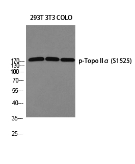
- Products Images

- Human lung squamous cell carcinoma tissue was stained with Anti-Topoisomerase IIα (ABT272) Antibody
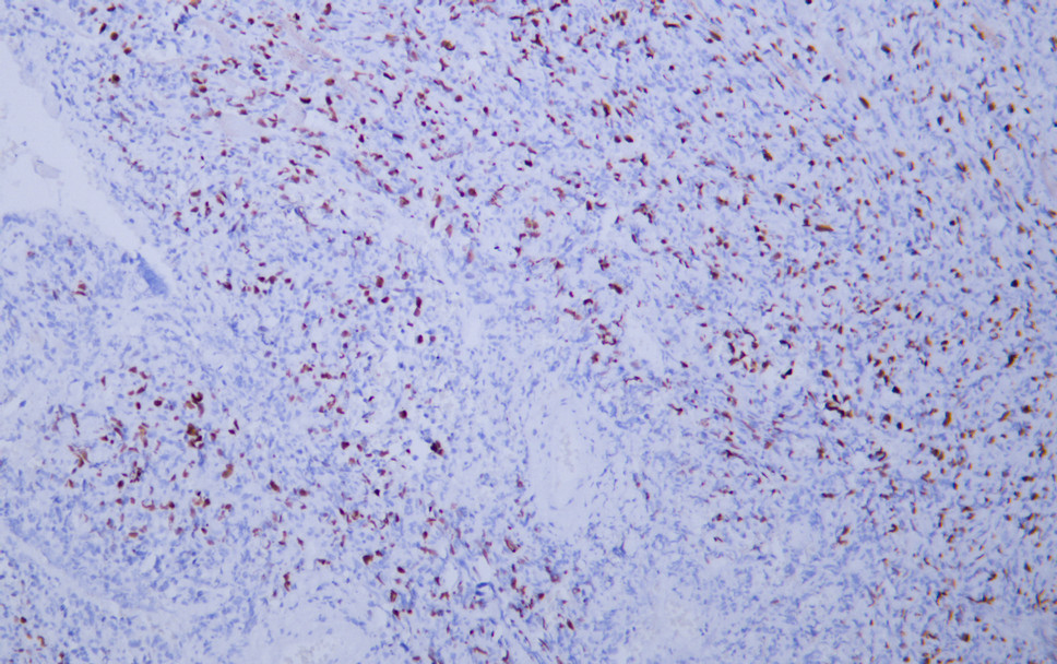
- Human lymphoma tissue was stained with Anti-Topoisomerase IIα (ABT272) Antibody
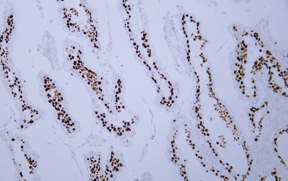
- Human seminoma tissue was stained with Anti-Topoisomerase IIα (ABT272) Antibody
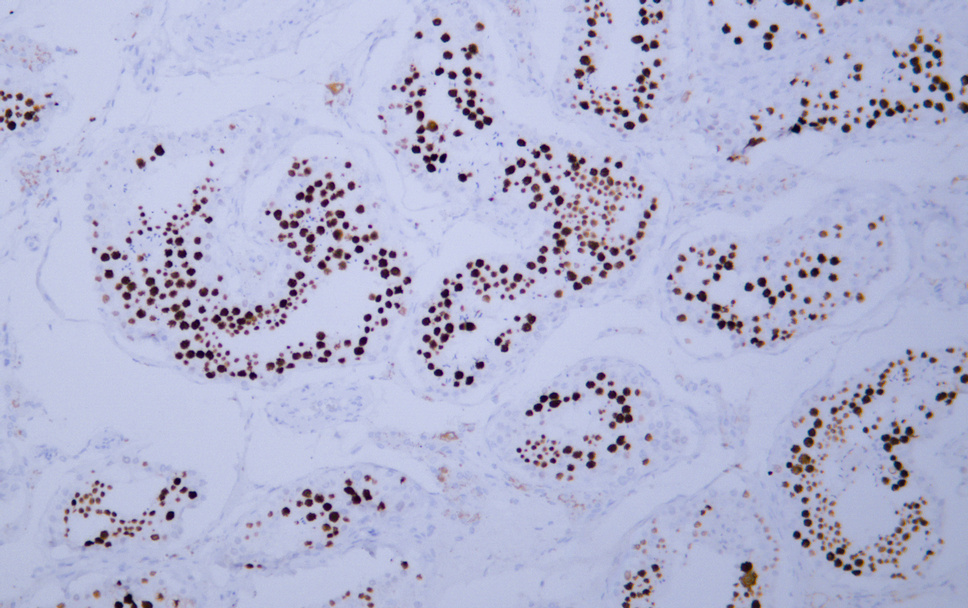
- Human testis tissue was stained with Anti-Topoisomerase IIα (ABT272) Antibody
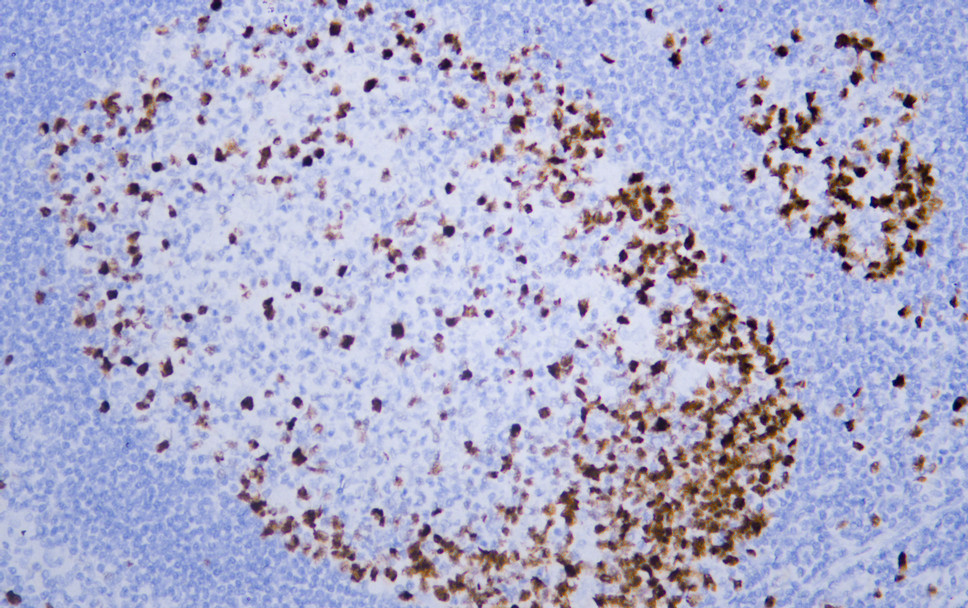
- Human tonsil tissue was stained with Anti-Topoisomerase IIα (ABT272) Antibody
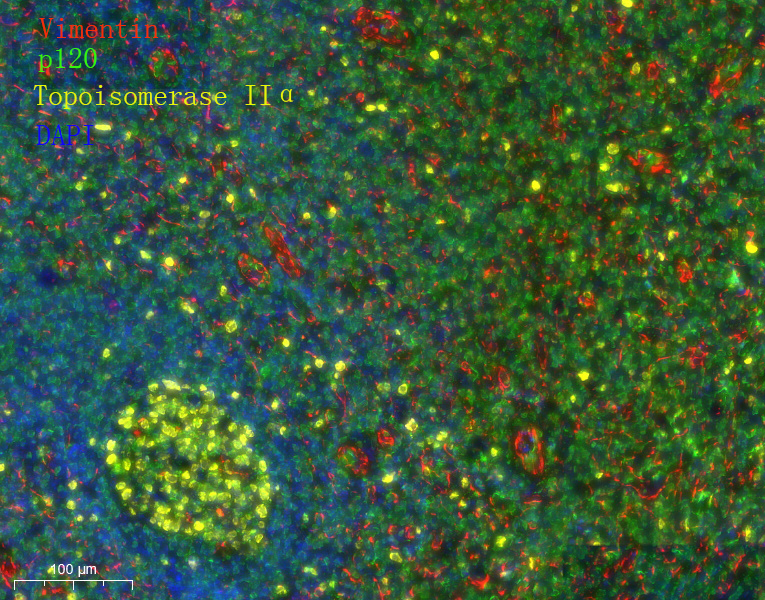
- Fluorescence multiplex immunohistochemical analysis of Human tonsil tissue (formalin-fixed paraffin-embedded section). Merged staining of Anti-Vimentin (YM6918), Anti-p120 (YM6086), Anti-Topoisomerase IIα (YM6914). The immunostaining was performed on a Leica Biosystems BOND® MAX instrument with an Sextuple-Fluorescence kit (RS0039, Immunoway). The section was incubated in 3 rounds of staining; sequentially for Anti-Vimentin (YM6918 1:200), Anti-p120 (YM6086 1:200), Anti-Topoisomerase IIα (YM6914 1:200).; each using a separate fluorescent tyramide signal amplification system. EDTA based antigen retrieval (Leica Biosystems BOND® Epitope Retrieval Solution 2, pH 9.0, 20 minutes) was used in between rounds of tyramide signal amplification to remove the antibody from the previous round, to avoid any cross-reactivity. DAPI (dark blue) was used as a nuclear counter stain. Microscopy and pseudocoloring of individual dyes was performed using a Slideviewer Imaging System (3D histech).

- Fluorescence multiplex immunohistochemical analysis of Human tonsil tissue (formalin-fixed paraffin-embedded section). Merged staining of Anti-Vimentin (YM6918), Anti-p120 (YM6086), Anti-Topoisomerase IIα (YM6914). The immunostaining was performed on a Leica Biosystems BOND® MAX instrument with an Sextuple-Fluorescence kit (RS0039, Immunoway). The section was incubated in 3 rounds of staining; sequentially for Anti-Vimentin (YM6918 1:200), Anti-p120 (YM6086 1:200), Anti-Topoisomerase IIα (YM6914 1:200).; each using a separate fluorescent tyramide signal amplification system. EDTA based antigen retrieval (Leica Biosystems BOND® Epitope Retrieval Solution 2, pH 9.0, 20 minutes) was used in between rounds of tyramide signal amplification to remove the antibody from the previous round, to avoid any cross-reactivity. DAPI (dark blue) was used as a nuclear counter stain. Microscopy and pseudocoloring of individual dyes was performed using a Slideviewer Imaging System (3D histech).