- 靶点:
- GLUT-1
- 简介:
- >>HIF-1 signaling pathway;>>Insulin secretion;>>Thyroid hormone signaling pathway;>>Adipocytokine signaling pathway;>>Glucagon signaling pathway;>>Insulin resistance;>>Bile secretion;>>Human T-cell leukemia virus 1 infection;>>Pathways in cancer;>>Renal cell carcinoma;>>Central carbon metabolism in cancer;>>Diabetic cardiomyopathy
- 基因名称:
- SLC2A1
- 蛋白名称:
- Solute carrier family 2 facilitated glucose transporter member 1
- Human Gene Id:
- 6513
- Human Swiss Prot No:
- P11166
- Mouse Gene Id:
- 20525
- Mouse Swiss Prot No:
- P17809
- Rat Gene Id:
- 24778
- Rat Swiss Prot No:
- P11167
- 免疫原:
- The antiserum was produced against synthesized peptide derived from human GLUT1. AA range:441-490
- 特异性:
- Glut1 Polyclonal Antibody detects endogenous levels of Glut1 protein.
- 组成:
- Liquid in PBS containing 50% glycerol, 0.5% BSA and 0.02% sodium azide.
- 来源:
- Polyclonal, Rabbit,IgG
- 稀释:
- IF 1:50-200 WB 1:500 - 1:2000. IHC 1:100 - 1:300. ELISA: 1:40000. Not yet tested in other applications.
- 纯化工艺:
- The antibody was affinity-purified from rabbit antiserum by affinity-chromatography using epitope-specific immunogen.
- 浓度:
- 1 mg/ml
- 储存:
- -15°C to -25°C/1 year(Do not lower than -25°C)
- 其他名称:
- SLC2A1;GLUT1;Solute carrier family 2; facilitated glucose transporter member 1;Glucose transporter type 1, erythrocyte/brain;GLUT-1;HepG2 glucose transporter
- 实测条带:
- 55kD
- 背景:
- This gene encodes a major glucose transporter in the mammalian blood-brain barrier. The encoded protein is found primarily in the cell membrane and on the cell surface, where it can also function as a receptor for human T-cell leukemia virus (HTLV) I and II. Mutations in this gene have been found in a family with paroxysmal exertion-induced dyskinesia. [provided by RefSeq, Apr 2013],
- 功能:
- disease:Defects in SLC2A1 are the cause of autosomal dominant GLUT1 deficiency syndrome [MIM:606777]; also called blood-brain barrier glucose transport defect. This disease causes a defect in glucose transport across the blood-brain barrier. It is characterized by infantile seizures, delayed development, and acquired microcephaly.,disease:Defects in SLC2A1 are the cause of dystonia type 18 (DYT18) [MIM:612126]. DYT18 is an exercise-induced paroxysmal dystonia/dyskinesia. Dystonia is defined by the presence of sustained involuntary muscle contraction, often leading to abnormal postures. DYT18 is characterized by attacks of involuntary movements triggered by certain stimuli such as sudden movement or prolonged exercise. In some patients involuntary exertion-induced dystonic, choreoathetotic, and ballistic movements may be associated with macrocytic hemolytic anemia.,function:Facilitative g
- 细胞定位:
- Cell membrane ; Multi-pass membrane protein . Melanosome . Photoreceptor inner segment . Localizes primarily at the cell surface (PubMed:18245775, PubMed:19449892, PubMed:23219802, PubMed:25982116, PubMed:24847886). Identified by mass spectrometry in melanosome fractions from stage I to stage IV (PubMed:17081065). .
- 组织表达:
- Detected in erythrocytes (at protein level). Expressed at variable levels in many human tissues.
CD38 affects the biological behavior and energy metabolism of nasopharyngeal carcinoma cells. INTERNATIONAL JOURNAL OF ONCOLOGY Int J Oncol. 2019 Feb;54(2):585-599 WB Human 1:1000 5-8F cell
货号:YT1928
Salidroside Induces Apoptosis in Human Gastric Cancer Cells via the Downregulation of ENO1/PKM2/GLUT1 Expression. BIOLOGICAL & PHARMACEUTICAL BULLETIN Biol Pharm Bull. 2021 Nov;44(11):1724-1731 IHC Mouse 1:100 MKN-45-Xenograft , SGC-7901 cell-Xenograft
货号:YT1928
Effect of IDH3a on glucose uptake in lung adenocarcinoma: A pilot study based on [18F]FDG. Cancer Medicine 2019 Jul 29 WB Human 1:500 A549 cell, H1299 cell
货号:yt1928
Loss of NDUFS1 promotes gastric cancer progression by activating the mitochondrial ROS-HIF1α-FBLN5 signaling pathway. BRITISH JOURNAL OF CANCER Jin Zhou WB Human 1:1000 MKN45 cell,N87 cell
货号:YT1928
Pancreatic Ductal Adenocarcinoma: The Characteristics of Contrast-Enhanced Ultrasound Are Correlated with the Hypoxic Microenvironment. Diagnostics Fang Nie IHC Human 1:200 pancreatic
货号:YT1928
Targeting blood brain barrier—Remote ischemic conditioning alleviates cognitive impairment in female APP/PS1 rats CNS Neuroscience & Therapeutics Yuxuan Ma IF,WB Rat 1:1000,1:800 cortex
货号:YT1928
Naringenin enhances the efficacy of ferroptosis inducers by attenuating aerobic glycolysis by activating the AMPK-PGC1α signalling axis in liver cancer Heliyon Yong-Zhuo Li WB Human HepG2 cell
货号:YT1928
- June 19-2018
- WESTERN IMMUNOBLOTTING PROTOCOL
- June 19-2018
- IMMUNOHISTOCHEMISTRY-PARAFFIN PROTOCOL
- June 19-2018
- IMMUNOFLUORESCENCE PROTOCOL
- September 08-2020
- FLOW-CYTOMEYRT-PROTOCOL
- May 20-2022
- Cell-Based ELISA│解您多样本WB检测之困扰
- July 13-2018
- CELL-BASED-ELISA-PROTOCOL-FOR-ACETYL-PROTEIN
- July 13-2018
- CELL-BASED-ELISA-PROTOCOL-FOR-PHOSPHO-PROTEIN
- July 13-2018
- Antibody-FAQs
- 产品图片

- Loss of NDUFS1 promotes gastric cancer progression by activating the mitochondrial ROS-HIF1α-FBLN5 signaling pathway. BRITISH JOURNAL OF CANCER Jin Zhou WB Human 1:5000 MKN45 cell,N87 cell,GES-1 cell,AGS cell,HGC-27 cell,KATO3 cell,SNU-1 cell
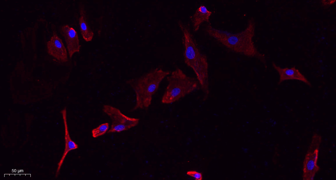
- Immunofluorescence analysis of A549. 1,primary Antibody(red) was diluted at 1:200(4°C overnight). 2, Goat Anti Rabbit IgG (H&L) - Alexa Fluor 594 Secondary antibody was diluted at 1:1000(room temperature, 50min).3, Picture B: DAPI(blue) 10min.
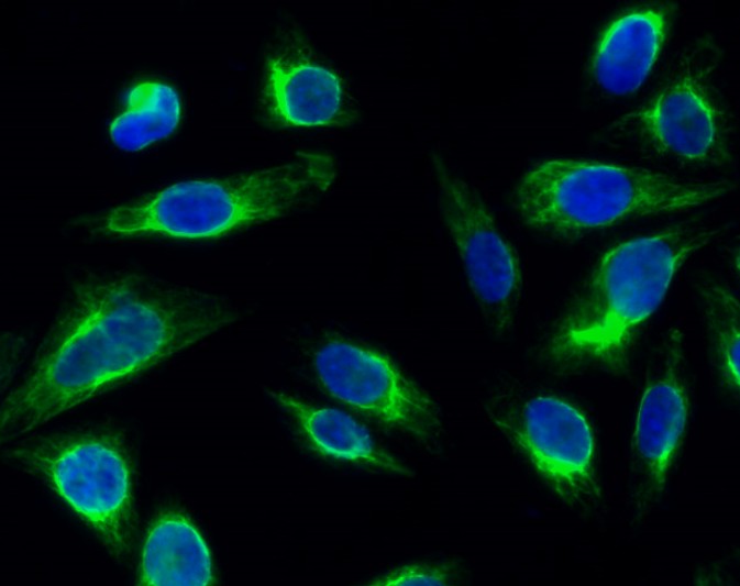
- Immunofluorescence analysis of Hela cell. 1,Glut1 Polyclonal Antibody(green) was diluted at 1:200(4° overnight). 2, Goat Anti Rabbit Alexa Fluor 488 Catalog:RS3211 was diluted at 1:1000(room temperature, 50min). 3 DAPI(blue) 10min.
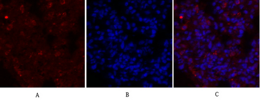
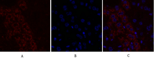
- Immunofluorescence analysis of rat-lung tissue. 1,Glut1 Polyclonal Antibody(red) was diluted at 1:200(4°C,overnight). 2, Cy3 labled Secondary antibody was diluted at 1:300(room temperature, 50min).3, Picture B: DAPI(blue) 10min. Picture A:Target. Picture B: DAPI. Picture C: merge of A+B
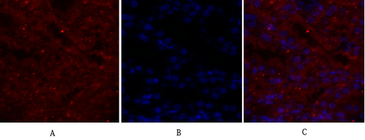
- Immunofluorescence analysis of rat-kidney tissue. 1,Glut1 Polyclonal Antibody(red) was diluted at 1:200(4°C,overnight). 2, Cy3 labled Secondary antibody was diluted at 1:300(room temperature, 50min).3, Picture B: DAPI(blue) 10min. Picture A:Target. Picture B: DAPI. Picture C: merge of A+B
- Immunofluorescence analysis of mouse-liver tissue. 1,Glut1 Polyclonal Antibody(red) was diluted at 1:200(4°C,overnight). 2, Cy3 labled Secondary antibody was diluted at 1:300(room temperature, 50min).3, Picture B: DAPI(blue) 10min. Picture A:Target. Picture B: DAPI. Picture C: merge of A+B
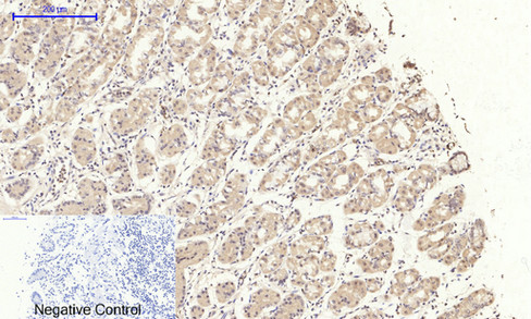
- Immunohistochemical analysis of paraffin-embedded Human-stomach tissue. 1,Glut1 Polyclonal Antibody was diluted at 1:200(4°C,overnight). 2, Sodium citrate pH 6.0 was used for antibody retrieval(>98°C,20min). 3,Secondary antibody was diluted at 1:200(room tempeRature, 30min). Negative control was used by secondary antibody only.
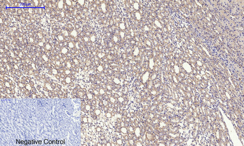
- Immunohistochemical analysis of paraffin-embedded Rat-kidney tissue. 1,Glut1 Polyclonal Antibody was diluted at 1:200(4°C,overnight). 2, Sodium citrate pH 6.0 was used for antibody retrieval(>98°C,20min). 3,Secondary antibody was diluted at 1:200(room tempeRature, 30min). Negative control was used by secondary antibody only.
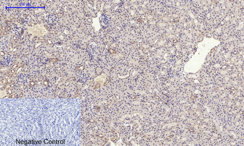
- Immunohistochemical analysis of paraffin-embedded Mouse-kidney tissue. 1,Glut1 Polyclonal Antibody was diluted at 1:200(4°C,overnight). 2, Sodium citrate pH 6.0 was used for antibody retrieval(>98°C,20min). 3,Secondary antibody was diluted at 1:200(room tempeRature, 30min). Negative control was used by secondary antibody only.
.jpg)
- Western Blot analysis of K562 cells using Glut1 Polyclonal Antibody diluted at 1:500

- Immunohistochemical analysis of paraffin-embedded Human placenta. 1, Antibody was diluted at 1:200(4° overnight). 2, High-pressure and temperature EDTA, pH8.0 was used for antigen retrieval. 3,Secondary antibody was diluted at 1:200(room temperature, 30min).
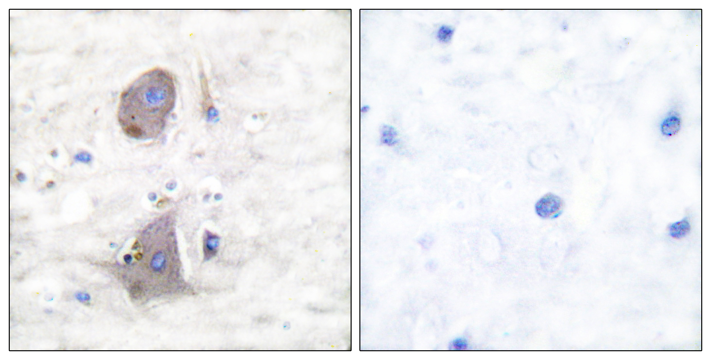
- Immunohistochemistry analysis of paraffin-embedded human brain tissue, using GLUT1 Antibody. The picture on the right is blocked with the synthesized peptide.

- Western blot analysis of lysates from Jurkat cells, using GLUT1 Antibody. The lane on the right is blocked with the synthesized peptide.
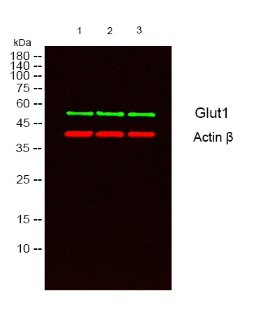
- Western blot analysis of lysates from 1)K562 , 2) Jurkat , 3) SW480 cells, (Green) primary antibody was diluted at 1:1000, 4°over night, secondary antibody(cat:RS23920)was diluted at 1:10000, 37° 1hour. (Red) Actin β Monoclonal Antibody(5B7) (cat:YM3028) antibody was diluted at 1:5000 as loading control, 4° over night,secondary antibody(cat:RS23710)was diluted at 1:10000, 37° 1hour.


