- 首页
- 公司介绍
- 热门促销
-
全部产品
-
试剂盒
- |
-
一抗
- |
-
二抗
- |
-
蛋白
- |
-
免疫组化试剂
- |
-
WB 试剂
- PonceauS Staining Solution
- PBST Washing Buffer, 10X
- 1.5M Tris-HCl Buffer, pH8.8
- 1M Tris-HCl Buffer, pH6.8
- 10% SDS Solution
- Prestained Protein Marker
- TBST Washing Buffer, 10X
- SDS PAGE Loading Buffer, 5X
- Stripping Buffered Solution
- Tris Buffer, pH7.4, 10X
- Total Protein Extraction Kit
- Running Buffer, 10X
- Transfer Buffer, 10X
- 30% Acr-Bis(29:1) Solution
- Tris电泳液速溶颗粒
- PBS(1X, premixed powder)
- TBS(1X, premixed powder)
- 快速封闭液
- 转膜液速溶颗粒
- Chemical reagents
- 公司新闻
- 营销网络
- 资源中心
- 联系我们
SDC3 rabbit pAb
- 货号:YT6720
- 应用:WB;IHC
- 种属:Human;Mouse;Rat
- 简介:
- >>Cell adhesion molecules
- 免疫原:
- Synthesized peptide derived from human SDC3 AA range: 131-181
- 特异性:
- This antibody detects endogenous levels of SDC3 at Human/Mouse/Rat
- 组成:
- Liquid in PBS containing 50% glycerol, 0.5% BSA and 0.02% sodium azide.
- 来源:
- Polyclonal, Rabbit,IgG
- 稀释:
- WB 1:500-2000;IHC 1:50-300
- 纯化工艺:
- The antibody was affinity-purified from rabbit antiserum by affinity-chromatography using epitope-specific immunogen.
- 储存:
- -15°C to -25°C/1 year(Do not lower than -25°C)
- 背景:
- The protein encoded by this gene belongs to the syndecan proteoglycan family. It may play a role in the organization of cell shape by affecting the actin cytoskeleton, possibly by transferring signals from the cell surface in a sugar-dependent mechanism. Allelic variants of this gene have been associated with obesity. [provided by RefSeq, Oct 2009],
- 功能:
- function:Cell surface proteoglycan that may bear heparan sulfate (By similarity). May have a role in the organization of cell shape by affecting the actin cytoskeleton, possibly by transferring signals from the cell surface in a sugar-dependent mechanism.,PTM:O-glycosylated within the Thr/Ser-rich region which could interact with lectin domains on other molecules.,similarity:Belongs to the syndecan proteoglycan family.,tissue specificity:Expressed in the nervous system, the adrenal gland, and the spleen.,
- 细胞定位:
- Cell membrane; Single-pass type I membrane protein.
- 组织表达:
- Expressed in the nervous system, the adrenal gland, and the spleen.
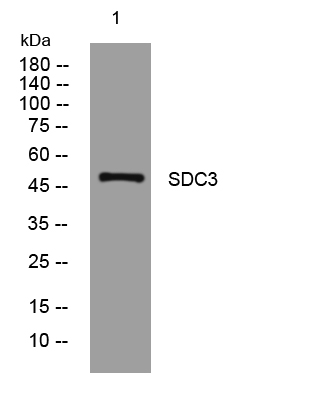
- Western blot analysis of lysates from HpeG2 cells, primary antibody was diluted at 1:1000, 4°over night
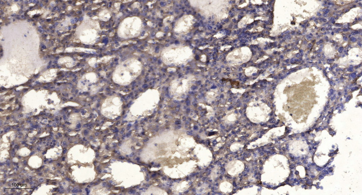
- Immunohistochemical analysis of paraffin-embedded human liver cancer. 1, Antibody was diluted at 1:200(4° overnight). 2, Tris-EDTA,pH9.0 was used for antigen retrieval. 3,Secondary antibody was diluted at 1:200(room temperature, 45min).


