VE-Cadherin Polyclonal Antibody
- 货号:YT5611
- 应用:IF;WB;IHC;ELISA
- 种属:Human;Mouse;Rat
- 简介:
- >>Cell adhesion molecules;>>Leukocyte transendothelial migration;>>Fluid shear stress and atherosclerosis
- 免疫原:
- The antiserum was produced against synthesized peptide derived from the Internal region of human CDH5. AA range:391-440
- 特异性:
- VE-Cadherin Polyclonal Antibody detects endogenous levels of VE-Cadherin protein.
- 组成:
- Liquid in PBS containing 50% glycerol, 0.5% BSA and 0.02% sodium azide.
- 来源:
- Polyclonal, Rabbit,IgG
- 稀释:
- IF 1:50-200 WB 1:500-2000, ELISA 1:10000-20000 IHC 1:50-300
- 纯化工艺:
- The antibody was affinity-purified from rabbit antiserum by affinity-chromatography using epitope-specific immunogen.
- 储存:
- -15°C to -25°C/1 year(Do not lower than -25°C)
- 其他名称:
- CDH5;Cadherin-5;7B4 antigen;Vascular endothelial cadherin;VE-cadherin;CD144
- 背景:
- This gene encodes a classical cadherin of the cadherin superfamily. The encoded preproprotein is proteolytically processed to generate the mature glycoprotein. This calcium-dependent cell-cell adhesion molecule is comprised of five extracellular cadherin repeats, a transmembrane region and a highly conserved cytoplasmic tail. Functioning as a classical cadherin by imparting to cells the ability to adhere in a homophilic manner, this protein plays a role in endothelial adherens junction assembly and maintenance. This gene is located in a gene cluster in a region on the long arm of chromosome 16 that is involved in loss of heterozygosity events in breast and prostate cancer. [provided by RefSeq, Nov 2015],
- 功能:
- function:Cadherins are calcium dependent cell adhesion proteins.,function:Cadherins are calcium dependent cell adhesion proteins. They preferentially interact with themselves in a homophilic manner in connecting cells; cadherins may thus contribute to the sorting of heterogeneous cell types. This cadherin may play a important role in endothelial cell biology through control of the cohesion and organization of the intercellular junctions. It associates with alpha-catenin forming a link to the cytoskeleton.,similarity:Contains 5 cadherin domains.,subcellular location:Found at cell-cell boundaries and probably at cell-matrix boundaries.,tissue specificity:Endothelial tissues and brain.,
- 细胞定位:
- Cell junction . Cell membrane ; Single-pass type I membrane protein . Found at cell-cell boundaries and probably at cell-matrix boundaries. KRIT1 and CDH5 reciprocally regulate their localization to endothelial cell-cell junctions. .
- 组织表达:
- Endothelial tissues and brain.
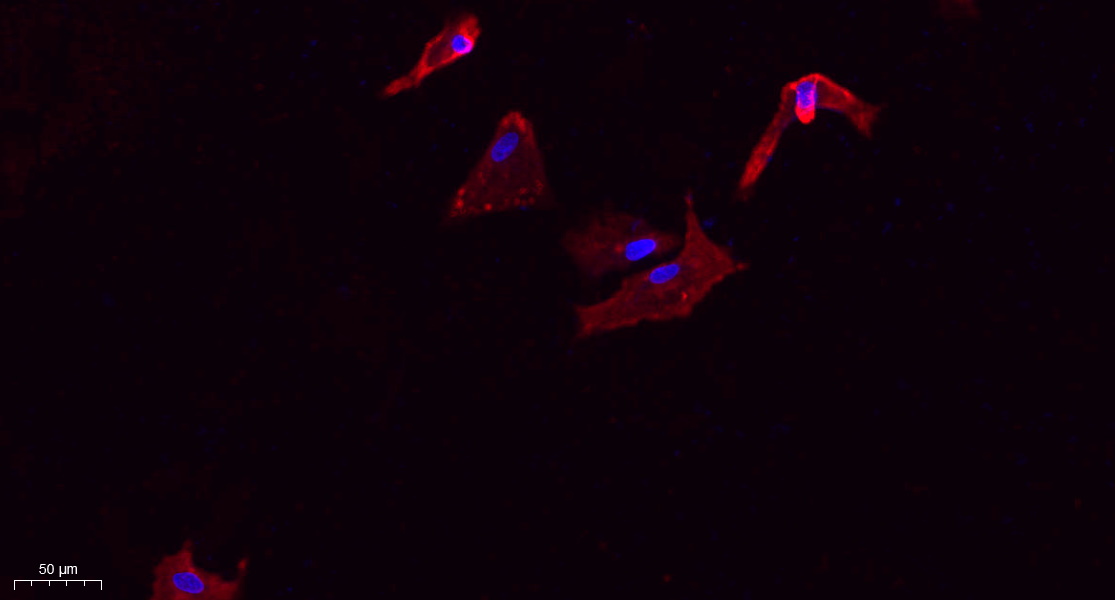
- Immunofluorescence analysis of A549. 1,primary Antibody(red) was diluted at 1:200(4°C overnight). 2, Goat Anti Rabbit IgG (H&L) - Alexa Fluor 594 Secondary antibody was diluted at 1:1000(room temperature, 50min).3, Picture B: DAPI(blue) 10min.
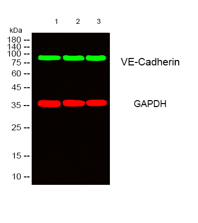
- Western blot analysis of lysates from 1) Hela, 2) mouse-lung ,3) mouse-kidney cells, (Green) primary antibody was diluted at 1:1000, 4°over night, secondary antibody(cat:RS23920)was diluted at 1:10000, 37° 1hour. (Red) GAPDH Monoclonal Antibody(2B8) (cat:YM3029) antibody was diluted at 1:5000 as loading control, 4° over night,secondary antibody(cat:RS23710)was diluted at 1:10000, 37° 1hour.
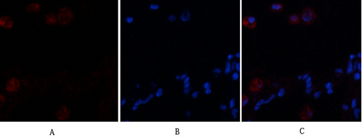
- Immunofluorescence analysis of human-lung tissue. 1,VE-Cadherin Polyclonal Antibody(red) was diluted at 1:200(4°C,overnight). 2, Cy3 labled Secondary antibody was diluted at 1:300(room temperature, 50min).3, Picture B: DAPI(blue) 10min. Picture A:Target. Picture B: DAPI. Picture C: merge of A+B

- Immunofluorescence analysis of rat-lung tissue. 1,VE-Cadherin Polyclonal Antibody(red) was diluted at 1:200(4°C,overnight). 2, Cy3 labled Secondary antibody was diluted at 1:300(room temperature, 50min).3, Picture B: DAPI(blue) 10min. Picture A:Target. Picture B: DAPI. Picture C: merge of A+B
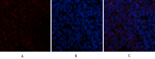
- Immunofluorescence analysis of rat-spleen tissue. 1,VE-Cadherin Polyclonal Antibody(red) was diluted at 1:200(4°C,overnight). 2, Cy3 labled Secondary antibody was diluted at 1:300(room temperature, 50min).3, Picture B: DAPI(blue) 10min. Picture A:Target. Picture B: DAPI. Picture C: merge of A+B
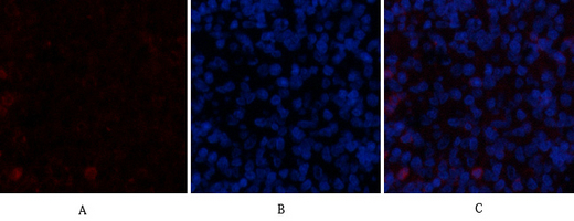
- Immunofluorescence analysis of rat-spleen tissue. 1,VE-Cadherin Polyclonal Antibody(red) was diluted at 1:200(4°C,overnight). 2, Cy3 labled Secondary antibody was diluted at 1:300(room temperature, 50min).3, Picture B: DAPI(blue) 10min. Picture A:Target. Picture B: DAPI. Picture C: merge of A+B
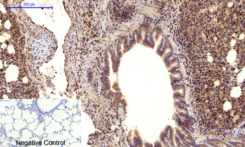
- Immunohistochemical analysis of paraffin-embedded Rat-lung tissue. 1,VE-Cadherin Polyclonal Antibody was diluted at 1:200(4°C,overnight). 2, Sodium citrate pH 6.0 was used for antibody retrieval(>98°C,20min). 3,Secondary antibody was diluted at 1:200(room tempeRature, 30min). Negative control was used by secondary antibody only.

- Immunohistochemical analysis of paraffin-embedded Rat-spleen tissue. 1,VE-Cadherin Polyclonal Antibody was diluted at 1:200(4°C,overnight). 2, Sodium citrate pH 6.0 was used for antibody retrieval(>98°C,20min). 3,Secondary antibody was diluted at 1:200(room tempeRature, 30min). Negative control was used by secondary antibody only.

- Western Blot analysis of Hela cells using VE-Cadherin Polyclonal Antibody. Antibody was diluted at 1:500. Secondary antibody(catalog#:RS0002) was diluted at 1:20000
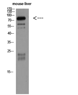
- Western Blot analysis of mouse-liver using VE-Cadherin Polyclonal Antibody diluted at 1:500. Secondary antibody(catalog#:RS0002) was diluted at 1:20000
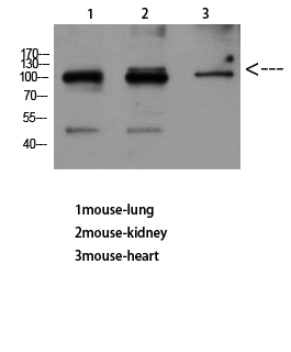
- Western Blot analysis of mouse-lung mouse-kidney mouse-heart using VE-Cadherin Polyclonal Antibody diluted at 1:500. Secondary antibody(catalog#:RS0002) was diluted at 1:20000