- 靶点:
- CD166
- 简介:
- >>Cell adhesion molecules
- 基因名称:
- ALCAM
- 蛋白名称:
- CD166 antigen
- Human Gene Id:
- 214
- Human Swiss Prot No:
- Q13740
- Mouse Gene Id:
- 11658
- Mouse Swiss Prot No:
- Q61490
- Rat Gene Id:
- 79559
- Rat Swiss Prot No:
- O35112
- 免疫原:
- The antiserum was produced against synthesized peptide derived from the C-terminal region of human ALCAM. AA range:481-530
- 特异性:
- CD166 Polyclonal Antibody detects endogenous levels of CD166 protein.
- 组成:
- Liquid in PBS containing 50% glycerol, 0.5% BSA and 0.02% sodium azide.
- 来源:
- Polyclonal, Rabbit,IgG
- 稀释:
- IF 1:50-200 WB 1:500 - 1:2000. ELISA: 1:20000. Not yet tested in other applications.
- 纯化工艺:
- The antibody was affinity-purified from rabbit antiserum by affinity-chromatography using epitope-specific immunogen.
- 浓度:
- 1 mg/ml
- 储存:
- -15°C to -25°C/1 year(Do not lower than -25°C)
- 其他名称:
- ALCAM;MEMD;CD166 antigen;Activated leukocyte cell adhesion molecule;CD166
- 实测条带:
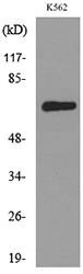
- 65kD
- 背景:
- This gene encodes activated leukocyte cell adhesion molecule (ALCAM), also known as CD166 (cluster of differentiation 166), which is a member of a subfamily of immunoglobulin receptors with five immunoglobulin-like domains (VVC2C2C2) in the extracellular domain. This protein binds to T-cell differentiation antigene CD6, and is implicated in the processes of cell adhesion and migration. Multiple alternatively spliced transcript variants encoding different isoforms have been found. [provided by RefSeq, Aug 2011],
- 功能:
- domain:The CD6 binding site is located in the N-terminal Ig-like domain.,function:Cell adhesion molecule that binds to CD6. Involved in neurite extension by neurons via heterophilic and homophilic interactions. May play a role in the binding of T- and B-cells to activated leukocytes, as well as in interactions between cells of the nervous system.,similarity:Contains 2 Ig-like V-type (immunoglobulin-like) domains.,similarity:Contains 3 Ig-like C2-type (immunoglobulin-like) domains.,tissue specificity:Spleen, placenta, liver, and weakly in liver. Expressed by activated T-cells, B-cells, monocytes and thymic epithelial cells. Expressed by neurons in the brain. Restricted expression in tumor cell lines. Preferentially expressed in highly metastasizing melanoma cell lines.,
- 细胞定位:
- Cell membrane ; Single-pass type I membrane protein . Cell projection, axon . Cell projection, dendrite . Detected at the immunological synapse, i.e, at the contact zone between antigen-presenting dendritic cells and T-cells (PubMed:15294938, PubMed:16352806). Colocalizes with CD6 and the TCR/CD3 complex at the immunological synapse (PubMed:15294938). .; [Isoform 3]: Secreted .
- 组织表达:
- Detected on hematopoietic stem cells derived from umbilical cord blood (PubMed:24740813). Detected on lymph vessel endothelial cells, skin and tonsil (PubMed:23169771). Detected on peripheral blood monocytes (PubMed:15048703). Detected on monocyte-derived dendritic cells (at protein level) (PubMed:16352806). Detected at low levels in spleen, placenta, liver (PubMed:9502422). Expressed by activated T-cells, B-cells, monocytes and thymic epithelial cells (PubMed:7760007). Isoform 1 and isoform 3 are detected in vein and artery endothelial cells, astrocytes, keratinocytes and artery smooth muscle cells (PubMed:15496415). Expressed by neurons in the brain. Restricted expression in tumor cell lines. Detected in highly metastasizing melanoma cell lines (PubMed:9502422).
A static magnetic field enhances the repair of osteoarthritic cartilage by promoting the migration of stem cells and chondrogenesis Journal of Orthopaedic Translation Ping Zhang IF Mouse knee joint
货号:YT5396
- June 19-2018
- WESTERN IMMUNOBLOTTING PROTOCOL
- June 19-2018
- IMMUNOHISTOCHEMISTRY-PARAFFIN PROTOCOL
- June 19-2018
- IMMUNOFLUORESCENCE PROTOCOL
- September 08-2020
- FLOW-CYTOMEYRT-PROTOCOL
- May 20-2022
- Cell-Based ELISA│解您多样本WB检测之困扰
- July 13-2018
- CELL-BASED-ELISA-PROTOCOL-FOR-ACETYL-PROTEIN
- July 13-2018
- CELL-BASED-ELISA-PROTOCOL-FOR-PHOSPHO-PROTEIN
- July 13-2018
- Antibody-FAQs
- 产品图片
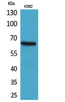
- Western Blot analysis of K562 cells using CD166 Polyclonal Antibody. Secondary antibody(catalog#:RS0002) was diluted at 1:20000
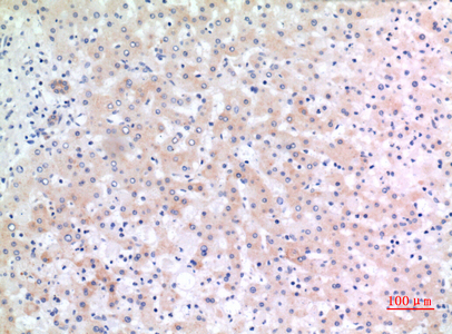
- Immunohistochemical analysis of paraffin-embedded human-liver, antibody was diluted at 1:100
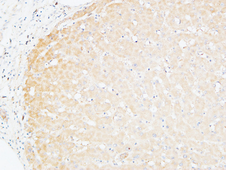
- Immunohistochemical analysis of paraffin-embedded Human Liver. 1, Antibody was diluted at 1:200(4° overnight). 2, High-pressure and temperature EDTA, pH8.0 was used for antigen retrieval. 3,Secondary antibody was diluted at 1:200(room temperature, 30min).
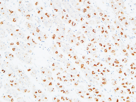
- Immunohistochemical analysis of paraffin-embedded Human stomach. 1, Antibody was diluted at 1:100(4° overnight). 2, High-pressure and temperature EDTA, pH8.0 was used for antigen retrieval. 3,Secondary antibody was diluted at 1:200(room temperature, 30min).
- Western blot analysis of lysate from K562 cells, using ALCAM Antibody.


